
Page 29 - BH-2-1
P. 29
Brain & Heart Pictorial rendition pulmonary stenosis
A B
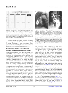
Figure 41. The incidence of abnormalities in interventricular septal Figure 42. Balloon pulmonary valvuloplasty (BPV) is likely to alleviate
motion following balloon pulmonary valvuloplasty (BPV). The prevalence pulmonary valve stenosis. In the absence of additional subvalvar or
of flat septal motion did not increase (P > 0.1) immediately after BPV (Pre supravalvar obstruction, the pulmonary circuit is subjected to increased
vs. Post BPV), nor did it increase (P > 0.1) at follow-up intermediate-term pulmonary arterial pressure and flow since most cyanotic heart defects
(ITFU). However, at follow-up long- term (LTFU), the prevalence of flat have either a large ventricular septal defect or a single ventricle. To
septal motion increased (P < 0.05), although this was in a small fraction circumvent this problem, there should be two or more obstructions to
of the total patients. Reproduced from Rao et al. 21 the pulmonary outflow tract before embarking on BPV for this group of
patients. 19,65 The cineangiographic frames above illustrate the presence of
subvalvar stenosis (filled white arrow [A] and filled lower black arrow
Long-term follow-up (defined as mean or median [B]), which are prerequisites to performing BPV. BPV will reduce/abolish
greater the 5 years) results of BPV have been investigated the gradient across the pulmonary valve, but the remaining subvalvar or
19,77,78
by several investigators. 68-75 These investigations reveal supravalvular stenosis will prevent flooding of the lungs. Reproduced
78
from Rao et al.
persistent effective relief of pulmonary valve obstruction
along with the development of varying degrees of PI. 68-75 held in Vienna, Austria, in February of 1987. At the
3. Pulmonary stenosis associated with conclusion of the presentation, Dr. Michael Tynan, the
chairman of the abstract session, congratulated the author
cyanotic congenital heart defects (CHDs) and colleagues with thanks for introducing an additional
Following the practice of using BPV for patients with indication for BPV. Thereafter, the author and colleagues,
isolated PS, as described in the preceding sections, along with other cardiologists, used this technique
the author and colleagues encountered an infant with to increase blood flow to the lungs as a substitute for a
d-transposition of the great vessels, a large ventricular Blalock-Taussig shunt, effectively relieving pulmonary
septal defect (VSD), severe subvalvar and valvar PS, and hypoperfusion and systemic arterial desaturation, as
19
77
19
hypoplastic pulmonary arteries. The data on this infant reviewed in the Rao, Rao and Brais publications. Rao,
78
77
were presented to the surgeons who worked with the Rao and Brais, Rao et al., suggested balloon sizes
author with a recommendation for an aortopulmonary slightly larger than pulmonary valve annulus diameter
shunt. The surgerical colleagues hesitated to surgically and require two or more obstructive elements in series
intervene because the aortopulmonary shunt was likely (as demonstrated in Figures 42-44) to avoid flooding of
to thrombose, given the diminished pulmonary arterial the lungs.
size. The following day, the infant was returned to the Two cohorts of patients were studied by the author: the
catheterization suite and underwent BPV. The procedure first consisted of eight infants, 76,77 and the second included
resulted in an increase in arterial oxygen saturation. fourteen patients. The most common diagnoses were (i)
78
Follow-up re-evaluation revealed good pulmonary arterial Fallot’s tetralogy and (ii) transposed great vessels with VSD
growth, and sometimes thereafter, the infant underwent and subvalvar and valvar PS. Peak pressure differences across
successful surgical correction of the defect. Based on the pulmonary valve were either eliminated or lowered after
this experience, the author and colleagues applied this BPV, but the subvalvar gradient remained (Figure 45). 77,78
technique to other infants needing palliation of pulmonary An increase in O2 saturation in the systemic circuit from
oligemia. Rao made a presentation demonstrating the 69.9 ± 11.5% to 81.4 ± 12.3 % (P < 0.05) (Figure 46),
76
utility of BPV in cyanotic heart disease patients at the indexed blood flow to the lungs from 1.83 ± 0.55 to
Pediatric Cardiology International Congress Conference 3.15 ± 1.38 l/min/m (P < 0.05) (Figure 47), pulmonary to
2
Volume 2 Issue 1 (2024) 13 https://doi.org/10.36922/bh.2406