
Page 110 - EJMO-9-2
P. 110
Eurasian Journal of
Medicine and Oncology UGVAE of breast lesions
A B C
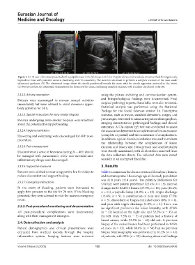
Figure 1. An 18-year-old woman presented with a palpable mass in the left breast. (A) Color Doppler ultrasound revealed a circumscribed, homogeneously
hypoechoic mass with posterior acoustic shadowing and rim vascularity. The decision was made to perform a complete excision of the mass under
ultrasound guidance. (B) The ultrasound image shows the needle positioned beneath the mass, with the needle apparatus centered on the lesion.
(C) Post-procedure, the ultrasound demonstrates the absence of the mass, confirming complete excision, with a marker clip placed at the site.
2.3.2.2. Activity resumption using the picture archiving and communication system,
Patients were encouraged to resume normal activities and histopathological findings were documented. Final
immediately but were advised to avoid strenuous upper- surgical pathology reports, if available, were also reviewed.
body activities for 24 h. Statistical analysis was performed using the Statistical
Package for the Social Sciences version 18. Descriptive
2.3.2.3. Special instructions for retro-areolar biopsies statistics, such as means, standard deviations, ranges, and
Patients undergoing retro-areolar biopsies were informed percentages, were used to summarize patient demographics,
about the potential for nipple bleeding. imaging characteristics, pathological findings, and clinical
outcomes. A Chi-square (χ ) test was conducted to assess
2
2.3.2.4. Hygiene restrictions the association between the completeness of lesion excision
Showering and swimming were discouraged for 48 h post- (complete or partial) and the occurrence of complications.
procedure. In addition, a point-biserial correlation was used to evaluate
the relationship between the completeness of lesion
2.3.2.5. Pain management excision and lesion size. Data privacy and confidentiality
Discomfort or a sense of heaviness lasting 24 – 48 h should were strictly maintained, with all identifiers removed from
be managed with paracetamol, while non-steroidal anti- the data collection sheets. The collected data were stored
inflammatory drugs were discouraged. securely in an encrypted Excel file.
2.3.2.6. Supportive measures 3. Results
Patients were advised to wear a supportive bra for 2 days to Table 1 summarizes the characteristics of the subject, lesions,
reduce discomfort and support healing. and mammograms. The average age of the study population
was 41.8 years (±14 years). The primary indications for
2.3.2.7. Emergency instructions
UGVAE were patient preference (22.4%; n = 15), interval
In the event of bleeding, patients were instructed to changes in BI-RADS 3 lesions (17.9%; n = 12), pain (16.4%;
apply firm pressure to the site for 20 min. If the bleeding n = 11), a palpable lump (14.9%; n = 10), nipple discharge
persisted, they were advised to visit the nearest emergency (13.4%; n = 9), a combination of pain and lump (7.5%;
room. n = 5), discordant or biopsy-indicated cases (6%; n = 4),
and pain with nipple discharge (1.5%; n = 1). There was
2.3.3. Post-procedural monitoring and documentation no significant preference for lesion laterality, with 47.8%
All post-procedural complications were documented, (n = 32) located on the right side and 52.2% (n = 35) on
along with their management strategies. the left. Only 7.5% (n = 5) of patients had a history of
breast cancer, while 92.5% (n = 62) did not. A previous
2.4. Data collection and analysis biopsy of the excised lesion had been performed in 19.5%
Patient demographics and clinical presentations were of cases (n = 13), while 80.6% (n = 54) had no previous
extracted from medical records through the hospital biopsy. Mammography was performed in 61.2% (n = 41)
information system. Imaging features were reviewed of patients, with 95% (n = 39) showing abnormal findings
Volume 9 Issue 2 (2025) 102 doi: 10.36922/ejmo.8436