
Page 210 - EJMO-9-2
P. 210
Eurasian Journal of
Medicine and Oncology Tetramethyl thyroxine boosts bladder cancer
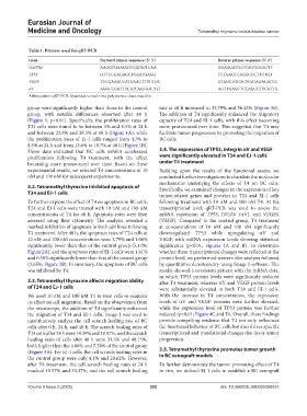
Table 1. Primers used for qRT‑PCR
Gene Forward primer sequence (5’‑3’) Reverse primer sequence (5’‑3’)
GAPDH AAGGTGAAGGTCGGAGTCAA GGAAGATGGTGATGGGATTT
TP53 GTTCCGAGAGCTGAATGAGG TCTGAGTCAGGCCCTTCTGT
VEGF TCCGAAACCATGAACTTTCTGC GTAGCTGCGCTGATAGACATCC
αV AGGCTGATTTCATCGGGGTTGT AGTTGAGTTCCAGCCTTCATTG
Abbreviation: qRT-PCR: Quantitative real-time polymerase chain reaction.
group were significantly higher than those in the control rate at 48 h increased to 33.79% and 56.42% (Figure 3B).
group, with notable differences observed after 48 h The addition of T4 significantly enhanced the migratory
(Figure 1, p<0.01). Specifically, the proliferation rates of capacity of T24 and EJ-1 cells, with this effect becoming
T24 cells were found to be between 5% and 8.5% at 24 h more pronounced over time. This suggested that T4 may
and between 22.8% and 28.3% at 48 h (Figure 1A), while facilitate tumor progression by promoting the migration of
the proliferation rates of EJ-1 cells ranged from 4.7% to BC cells.
8.5% at 24 h and from 13.6% to 18.7% at 48 h (Figure 1B).
These data indicated that BC cells exhibit accelerated 3.4. The expression of TP53, integrin αV and VEGF
proliferation following T4 treatment, with the effect were significantly elevated in T24 and EJ-1 cells
becoming more pronounced over time. Based on these under T4 treatment
experimental results, we selected T4 concentrations of 10 Building upon the results of the functional assays, we
nM and 100 nM for subsequent experiments. conducted further investigations to elucidate the molecular
mechanisms underlying the effects of T4 on BC cells.
3.2. Tetramethyl thyroxine inhibited apoptosis of Specifically, we examined changes in the expression of key
T24 and EJ-1 cells tumor-related genes and proteins in T24 and EJ-1 cells
To further explore the effect of T4 on apoptosis in BC cells, following treatment with 10 nM and 100 nM T4. At the
T24 and EJ-1 cells were treated with 10 nM and 100 nM transcriptional level, qRT-PCR was used to assess the
concentrations of T4 for 48 h. Apoptotic rates were then mRNA expression of TP53, ITGAV (αV), and VEGFA
assessed using flow cytometry. The analysis revealed a (VEGF). Compared to the control group, T4 treatment
marked inhibition of apoptosis in both cell lines following at concentrations of 10 nM and 100 nM significantly
T4 treatment. After 48 h, the apoptosis rates of T24 cells at downregulated TP53 while upregulating αV and
10 nM and 100 nM concentrations were 2.79% and 1.66% VEGF, with mRNA expression levels showing statistical
significantly lower than that of the control group (5.14%; significance (p<0.01; Figures 4A and B). To determine
Figure 2A), and the apoptosis rates of EJ-1 cells were 1.81% whether these transcriptional changes were reflected at the
and 0.58% significantly lower than that of the control group protein level, we performed western blot analysis followed
(3.65%; Figure 2B). In summary, the apoptosis of BC cells by quantitative densitometry using Image J software. The
was inhibited by T4. results showed a consistent pattern with the mRNA data,
in which TP53 protein levels were significantly reduced
3.3. Tetramethyl thyroxine affects migration ability after T4 treatment, whereas αV and VEGF protein levels
of T24 and EJ-1 cells were substantially elevated in both T24 and EJ-1 cells.
We used 10 nM and 100 nM T4 to treat cells to examine With the increase in T4 concentration, the expression
its effect on cell migration. Based on the observation from levels of αV and VEGF proteins were further elevated,
the microscope, the addition of T4 significantly enhanced while the expression level of TP53 protein was further
the migration of T24 and EJ-1 cells. Image J was used to reduced (p<0.01; Figure 4C and D). Overall, these findings
quantitatively analyze the cell scratch healing rate of BC provide compelling evidence that T4 not only influences
cells after 0 h, 24 h, and 48 h. The scratch healing rates of the functional behavior of BC cells but also drives specific
T24 cells after 24 h were 14.39% and 19.97%, and the scratch transcriptional and translational changes that favor tumor
healing rates of cells after 48 h were 31.5% and 48.75%, progression.
both higher than the 4.66% and 7.74% of the control group
(Figure 3A). For EJ-1 cells, the cell scratch healing rates in 3.5. Tetramethyl thyroxine promotes tumor growth
the control group were only 4.1% and 20.62%. However, in BC xenograft models
after T4 treatment, the cell scratch healing rates at 24 h To further demonstrate the tumor-promoting effect of T4
reached 15.57% and 33.37%, and the cell scratch healing in vivo, we utilized EJ-1 cells to establish a BC xenograft
Volume 9 Issue 2 (2025) 202 doi: 10.36922/EJMO025080037