
Page 211 - EJMO-9-2
P. 211
Eurasian Journal of
Medicine and Oncology Tetramethyl thyroxine boosts bladder cancer
A tumor model. Tumor-bearing mice were divided into a
control group and a T4 treatment group, with five mice
in each group. Changes in tumor volume were recorded
every 3 days. By the 30 day, the tumor volume in the
th
T4 treatment group reached 545.47 mm³, compared to
251.5 mm³ in the control group (Figure 5A and B). On
th
the 30 day, the mice were sacrificed, and the tumors
were excised and weighed. The mass of the tumor in the
T4 group post-treatment (0.58044 g) was significantly
higher than that in the untreated group (0.29566 g; p<0.01;
B
Figure 5A and C). In addition, we collected tail blood
samples from the mice to measure the concentrations
of T4, T3, and TSH in the serum, with T4 serving as the
precursor to T3. The observed increase in serum T4 and T3
levels, along with a decrease in TSH (Figure 5D), indicates
signs of hyperthyroidism. The experimental results
11
revealed that by the 28 day, the serum levels of T4 and
th
T3 had significantly increased, while the concentration of
TSH had decreased. T4-induced hyperthyroidism in mice
may influence the course of tumor development.
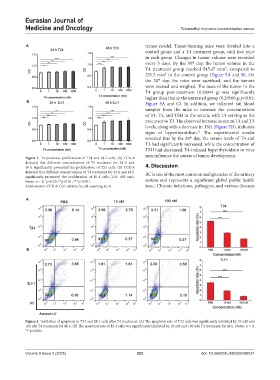
Figure 1. T4 promotes proliferation of T24 and EJ-1 cells. (A) CCK-8
detected that different concentrations of T4 treatment for 24 h and
48 h significantly promoted the proliferation of T24 cells. (B) CCK-8 4. Discussion
detected that different concentrations of T4 treatment for 24 h and 48 h BC is one of the most common malignancies of the urinary
significantly promoted the proliferation of EJ-1 cells (OD: 450 nm).
Notes: n = 3; *p<0.05; **p<0.01; ***p<0.001. system and represents a significant global public health
Abbreviation: CCK-8: Cell viability by cell counting kit-8. issue. Chronic infections, pathogens, and various diseases
A
B
Figure 2. Inhibition of apoptosis in T24 and EJ-1 cells after T4 treatment. (A) The apoptosis rate of T24 cells was significantly inhibited by 10 nM and
100 nM T4 treatment for 48 h. (B) The apoptosis rate of EJ-1 cells was significantly inhibited by 10 nM and 100 nM T4 treatment for 48 h. Notes: n = 3;
***p<0.001.
Volume 9 Issue 2 (2025) 203 doi: 10.36922/EJMO025080037