
Page 83 - ESAM-1-4
P. 83
Engineering Science in
Additive Manufacturing TwinPrint: Dual-arm robotic bioprinting
cells (BM-MSCs). The HL60 and BM-MSCs cell lines were Confocal Microscope, Germany) was used to observe
supplied by ATCC (USA) and Professor Abdalla Awidi and evaluate cells within the printed constructs. Human
(Cell Therapy Center, University of Jordan, Amman, BM-MSCs stained with CMFDA Dye were observed using
Jordan), respectively. Human BM-MSCs were cultured 492 nm excitation and 517 nm emission filters. HL-60 cells
and maintained, as described before. Briefly, the cells were stained with 5-chloromethyl tetraphenyl-p-xylylene
2
3
cultured at a seeding density of 4×10 cells/cm ; when (CMTPX) dye were observed using 577 nm excitation and
cultures reached 80% confluence, the cells were subcultured 602 nm emission filters. Z-stack images were obtained to
using 0.25% trypsin. Cells in passages 4–8 were used in the evaluate the 3D distribution of cells within the printed cell-
bioprinting experiments. Human BM-MSCs cells were laden constructs.
maintained in α-modified minimum essential medium
(α-MEM) supplemented with 10% mesenchymal stem 3. Results and discussion
cell–qualified fetal bovine serum (FBS), 2 mM L-glutamine, 3.1. TwinPrint system
and 1% penicillin/streptomycin (GIBCO, ThermoFisher,
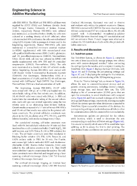
USA). HL-60 AML cell line was cultured in RPMI-1640 The TwinPrint System, as shown in Figure 2, comprises
media supplemented with 10% FBS and 1% penicillin/ two sets of four microfluidic syringe pumps, two robotic
23
streptomycin (GIBCO, ThermoFisher, USA). The cells arms with custom-designed nozzles, tubes connecting
were maintained at a density of 500×10 cells/mL media. the syringe tips to the nozzles, and a computer running the
3
Before bioprinting, human BM-MSCs were stained with TwinPrint GUI. Figure 3 displays the four GUI tabs, with
Cell Tracker Green 5-chloromethyl fluorescein diacetate Figure 3A and B showing the pre-printing settings and
™
(CMFDA) Dye (Invitrogen, ThermoFisher, USA) at a Figure 3C and D displaying the settings for the initiation,
final concentration of 10 µM, and the HL-60 cell line was control, and monitoring of the 3D bioprinting process.
stained with CellTracker Red CMTPX Dye (Invitrogen, First, the “Device Settings” tab, as shown in Figure 3A,
™
ThermoFisher, USA) at a final concentration of 5 µM. allows the user to connect/disconnect devices and set
For bioprinting, human BM-MSCs (3×10 cells) generic printing parameters, including desired z-height,
6
were mixed with 500 µL of 1× PBS and loaded into the pump syringe type, and desired flow rate. The GUI
microfluidic tubing of the first robotic arm. In addition, displays a list of communication (COM) ports that are
HL-60 (9×10 cells) were mixed with 500 µL 1× PBS and open for connection, to avoid interference with occupied
6
loaded into the microfluidic tubing of the second robotic ports. Figure 4A and B are zoomed-in images of the Dobot
settings and Pump settings, respectively, showing examples
arm. Each cell type was printed separately using the two
robotic arms in an alternating layer-by-layer fashion. of how the system operates when devices are connected to
Growth media composed of a 1:1 ratio of human BM-MSCs TwinPrint. Upon successful connection of the devices, the
and HL-60 cell line media was added to the printed cell- corresponding settings are activated across all tabs to allow
laden constructs. The printed cell-laden constructs were for communication with the connected devices exclusively.
placed in the CO incubator set at 37°C, 5% CO , and 95% Instantaneous updates are provided for the robotic
2
2
relative humidity, with media exchange every 3 days. arm’s location, which is used to determine the arm
corresponding to each box of Dobot settings (Figure 4A).
For cytoskeletal staining, cell-laden constructs were
fixed with 4% formaldehyde solution for 30 min and then It is crucial to identify which arm is R1 to avoid false layer
splitting, as R1 is always the first to start printing. The
incubated in a cold cytoskeleton buffer (3 mM MgCl , 300 user-defined home coordinates are used as a starting point
2
mM sucrose, and 0.5% Triton X-100 in PBS solution) for
5 min. The cell-laden constructs were then incubated in
blocking buffer solution (5% FBS, 0.1% Tween-20, and
0.02% sodium azide in PBS) for 30 min. For F-actin,
anti-mouse IgG (whole molecule)-FITC and rhodamine-
phalloidin (1:300; Thermo Fisher Scientific, USA) were
added to the cell-laden constructs for 1 h. Then DAPI
were added for 5 min to counterstain the nucleus. The cell-
laden constructs were observed and imaged using a laser
scanning confocal microscope (Zeiss LSM 710 Inverted
Confocal Microscope, Germany).
For confocal microscopy imaging, an inverted laser
scanning confocal microscope (Zeiss LSM 880 Inverted Figure 2. The TwinPrint system with an older version of the user interface
Volume 1 Issue 4 (2025) 6 doi: 10.36922/ESAM025410025