
Page 13 - GTM-1-1
P. 13
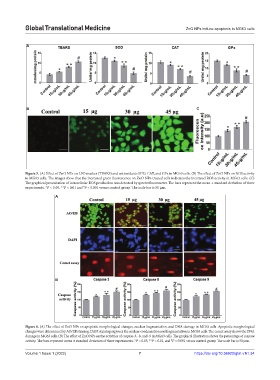
Global Translational Medicine                                       ZnO NPs induce apoptosis in MG63 cells


            A


            B                                                                        C


            Figure 5. (A) Effect of ZnO NPs on LPO marker (TBARS) and antioxidants SOD, CAT, and GPx in MG63 cells. (B) The effect of ZnO NPs on ROS activity
            in MG63 cells. The images show that the increased green fluorescence on ZnO NPs-treated cells indicates the increased ROS activity in MG63 cells. (C)
            The graphical presentation of intracellular ROS production was detected by spectrofluorometer. The bars represent the mean ± standard deviation of three
            experiments. *P < 0.05, **P < 0.01 and  P < 0.001 versus control group. The scale bar is 50 µm.
                                     #
                         A


                         B


            Figure 6. (A) The effect of ZnO NPs on apoptotic morphological changes, nuclear fragmentation, and DNA damage in MG63 cells. Apoptotic morphological
            changes were determined by AO/EB staining. DAPI staining explores the nuclear condensation and fragmentation in MG63 cells. The comet assay shows the DNA
            damage in MG63 cells. (B) The effect of ZnO NPs on the activities of caspase-3, -8, and -9 in MG63 cells. The graphical illustration shows the percentage of caspase
                                                                          #
            activity. The bars represent mean ± standard deviation of three experiments. *P < 0.05, **P < 0.01, and  P < 0.001 versus control group. The scale bar is 50 µm.
            Volume 1 Issue 1 (2022)                         7                       https://doi.org/10.36922/gtm.v1i1.34