
Page 14 - GTM-1-1
P. 14
Global Translational Medicine ZnO NPs induce apoptosis in MG63 cells
3.10. Effect of ZnO NPs on DNA damage apoptosis-related proteins in ZnO NPs-treated MG63 cells.
The comet test demonstrated that the ZnO NPs caused In comparison to untreated cells, the injection of ZnO
DNA damage in MG63 cells. In untreated control cells, NPs significantly reduced the expression levels of p-P13K,
we observed normal, intact DNA in the electrophoresis p-AKT, and p-mTOR, depending on the concentrations
plot. On the contrary, the damaged increased head DNA applied (Figure 8A and B). The activation of multiple
was detected in ZnO NPs-exposed cells. The increased conjugation processes mediate the lipidation of LC3 onto
DNA head DNA was directly associated with increasing cell membranes, thereby transforming LC3-I to LC3-II.
concentrations of ZnO NPs (Figure 6A). The LC3-positive puncta indicative of this lapidated form
is required for autophagosome formation. The treatment
3.11. Effect of ZnO NPs on caspase-3, -8, and -9 of ZnO NPs at the doses utilized in MG63 cells caused the
The expression of caspase-3, -8, and -9 in MG63 cells was conversion of LC3-I to its lapidated LC3-II form, as shown
ascertained by standard protocols. When comparing ZnO by Western blot results. Furthermore, ZnO NPs treatment
NPs-administered MG63 cells to control cells, the levels of considerably increased beclin1 expression. Nevertheless,
caspase-3, -8, and -9 were increased in a concentration- another autophagy marker, P62, was dramatically decreased
dependent manner. The graphical representation of by MG63 treatment at doses determined (Figure 8C). The
caspase-3, -8, and -9 is depicted graphically (Figure 6B). relative expression of LC3, beclin-1, and P62 versus β-actin
is depicted in the graph (Figure 8D).
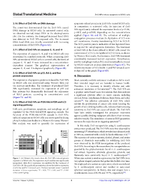
3.12. Effect of ZnO NPs on p53, Bcl-2, and Bax
protein expression 4. Discussion
The expression of apoptotic proteins induced by ZnO NPs Most currently available anticancer medications fail to reach
in MG63 cells was determined using Western blots and their intended target and are harmful to human health.
the relevant antibodies. The treatment of produced ZnO Therefore, it is necessary to identify effective and safer
NPs significantly increased the expression of p53 and anticancer medicines or formulations . The ZnO NPs are
[19]
Bax proteins but dramatically decreased the expression a popular metal-based nano-formulation that demonstrates
of Bcl-2 proteins, according to concentrations used a significant cytotoxic effect on many cancers, including
(Figure 7A and B). cervical, breast, lymphomas, leukemia, bone, brain, and colon
[20]
3.13. Effect of ZnO NPs on PI3K/Akt/mTOR and LC3 cancers . The selective cytotoxicity of ZnO NPs, which
signaling pathway inhibits the proliferation of cancer cells while boosting the
growth of non-dividing healthy cells, indicates that increasing
Cell cycle, proliferation, apoptosis, and autophagy are all sensitivity to ZnO NPs can cause cell death. The ZnO NPs
regulated by the PI3K/Akt/mTOR signaling cascade. The have been widely investigated for anticancer treatments
function of the PI3K/Akt/mTOR cascade in ZnO NPs- against quickly dividing malignant cells due to their intrinsic
induced apoptosis in MG63 cells was investigated utilizing selective toxicity. The stimulation of extreme ROS generation
phosphorylated antibodies in a Western blot assay. Western serves as the major factor in triggering apoptosis .
[21]
blot analysis was used to quantify the expression of
In this study, ZnO nanoparticles using S. xanthocarpum
extract were produced. The ZnO NPs were first observed by
A B measuring UV absorbance, which revealed an enhanced peak
at 380 nm, consistent with a study by Santhoshkumar et al.
[22]
The existence of various saponin, alcohol, phenol, and amine
groups of chemicals responsible for stabilizing ZnO NPs
were observed by the FTIR investigations of the produced
ZnO NPs. According to the researchers, the nanoparticles are
guarded by the phytochemicals found in S. xanthocarpum
leaf extract. Saponins, alkaloids, glycosides, phenols, and
flavonoids were commonly found in S. xanthocarpum .
[23]
Figure 7. (A) Western blot results show the ZnO NPs-induced protein When zinc acetate is used as a precursor, the ZnO NPs form
expression in MG63 cells. The expression of p53 and Bax was significantly tiny sphere-shaped particles that gather like bullets over time.
upregulated, while Bcl-2 expression was inhibited by the treatment of We employed zinc acetate as a precursor, which was reduced
ZnO NPs. (B) The graphical representation shows the relative expression
of p53, Bax and Bcl-2 versus β-actin. The bars represent mean ± standard by S. xanthocarpum extract and resulted in nano-sized
deviation of three experiments. *P < 0.05, **P < 0.01, and P < 0.001 spherical nanoparticles. In theory, zinc and oxygen have a
#
versus control group. stoichiometric mass of 80.3% and 19.7%, respectively .
[24]
Volume 1 Issue 1 (2022) 8 https://doi.org/10.36922/gtm.v1i1.34