
Page 21 - GTM-1-1
P. 21
Global Translational Medicine Anticancer effect of S. xanthocarpum on KB cell line
administering S. xanthocarpum extract in different doses dismutase (SOD) and catalase (CAT) activities were
(50 – 350 µg/ml), the cells were then incubated overnight in measured using the methods by Beyer and Fridovich
[14]
CO incubator. Afterward, MTT dye was added to each well and Aebi , respectively. The measurement of glutathione
[15]
2
in a series of doses (50, 100, 150, 200, 300, and 350 µg/ml) and (GSH) content was carried out using the method by
the cells were incubated for an additional 4 h at 37°C. After Griffith and 5,5’dithiobis-2- nitrobenzoic acid, DTNB
[16]
the brief incubation, the DMEM medium in each well was (Sigma chemical Co., USA). Measurement of MAD
discarded and DMSO was added to dissolve the formazan. formation was accomplished through its response with
The absorbance was measured at 490 nm in a microplate thiobarbituric acid reactive substances (TBARS) using the
reader (Bio-Rad). The absorbance data were expressed in method by Ohkawa et al. (1979) .
[17]
percentage with respect to the control. The half maximal
inhibitory concentration (IC ) values were calculated and 2.10. Statistical analysis
50
the optimum doses were analyzed at different time period. The data are expressed as mean ± standard deviation (SD)
and statistical comparisons were carried out by one-way
2.6. Measurement of ROS in KB cells analysis of variance and Duncan’s multiple range test using
DCF oxidized by radicals were visualized at excitation SPSS version 17.0. The results were considered statistically
wavelength of 535 nm and emission wavelength of 485 nm. significant if P < 0.05.
DCF is not oxidized by hydrogen peroxide or superoxide
anion radical. Overnight grown cells were treated in 24-well 3. Results
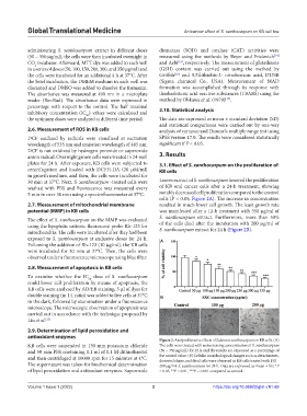
plates for 24 h. After exposure, KB cells were subjected to 3.1. Effect of S. xanthocarpum on the proliferation of
centrifugation and loaded with DCFH-DA (20 µM/ml) KB cells
in growth medium, and then, the cells were incubated for
30 min at 37°C. Next, S. xanthocarpum -treated cells were Leaves extract of S. xanthocarpum lowered the proliferation
washed with PBS and fluorescence was measured every of KB oral cancer cells after a 24-h treatment, showing
5 min in over 30 min using a spectrofluorometer at 37°C. notably decreased cell proliferation compared to the control
cells (P < 0.05; Figure 2A). The increase in concentration
2.7. Measurement of mitochondrial membrane resulted in much lower cell growth. The least growth rate
potential (MMP) in KB cells was manifested after a 12-h treatment with 350 µg/ml of
The effect of S. xanthocarpum on the MMP was evaluated S. xanthocarpum extract. Furthermore, more than 50%
using the lipophilic cationic fluorescent probe Rh-123 for of the cells died after the incubation with 200 µg/ml of
mitochondria. The cells were incubated after they had been S. xanthocarpum extract for 24 h (Figure 2B).
exposed to S. xanthocarpum at exclusive doses for 24 h. A
Following the addition of Rh-123 (10 µg/ml), the KB cells
were incubated for 30 min at 37°C. Then, the cells were
observed under a fluorescence microscope using blue filter.
2.8. Measurement of apoptosis in KB cells
To examine whether the IC dose of S. xanthocarpum
50
could lower cell proliferation by means of apoptosis, the
KB cells were analyzed by AO/EB staining, 5 µl of dyes for
double staining (in 1:1 ratio) was added to live cells at 37°C B
in the dark, followed by examination under a fluorescence
microscope. The microscopic observation of apoptosis was
carried out in accordance with the technique proposed by
Liu et al. [13]
2.9. Determination of lipid peroxidation and
antioxidant enzymes
Figure 2. Antiproliferative effects of Solanum xanthocarpum on KB cells. (A)
KB cells were suspended in 130 mm potassium chloride The cells were treated with an increasing concentration of S. xanthocarpum
and 50 mm PBS containing 0.1 ml of 0.1 M dithiothreitol (50 – 350 µg/mL) for 24 h and the results are expressed as a percentage of
and then centrifuged at 10000 rpm for 15 minutes at 4°C. the control value. (B) Cellular morphological changes such as detachments,
distorted shape, and dead cells were observed in KB cells treated with 150 –
The supernatant was taken for biochemical determination 200 µg/mL S. xanthocarpum for 24 h. Data are expressed as mean ± SD. *P
of lipid peroxidation and antixodant enzymes. Superoxide < 0.05, **P < 0.01, ***P < 0.001 compared to control.
Volume 1 Issue 1 (2022) 3 https://doi.org/10.36922/gtm.v1i1.68