
Page 111 - GTM-2-3
P. 111
Global Translational Medicine TEs link to Parkinson’s risk and progression
A
B C
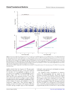
Figure 2. Association analysis identified the associations of TEs with the risk and the progression of PD. (A) Manhattan plot of TE-GWAS obtained from
the logistic regression model, comparing PD cases (n = 1,221) with healthy controls (n = 689). Each data point on the plot represents a TE. The Y-axis shows
the original P-values after the -log transformation of each TE association; the X-axis shows the sequential position of TEs by chromosome (not real scale).
10
The dashed red line corresponds to the significance threshold (FDR-adjusted P-value lower than 5%). (B) Adjusted mean Hoehn and Yahr stage across
time predicted by the TE-LMM model for PD cases with chr8_114592257_ALU carriers (magenta line) and non-carriers (light-blue line). (C) Adjusted
mean MDS-UPDRS I score across time predicted by the TE-LMM model for PD cases with chr13_81793576_SVA carriers (magenta line) and non-carriers
(light-blue line). Shaded ribbons indicate the standard error of the mean.
Abbreviations: TE-LMM: Transposable element-linear mixed model; TE: Transposable element; PD: Parkinson’s disease; TE-GWAS: TE Genome-Wide
Association Study.
analysis revealed that the 26 genes (27 TE–gene pairs) levels and a more pronounced involvement in immune-
affected by interaction-TE-eQTL cis loci were related to related signaling pathways.
the formation of immunoglobulin complexes and were We conducted further investigations to determine
involved in the recognition of immunoglobulin receptor whether TE polymorphisms have an impact on the
binding (Figure S5A). The 624 eGenes (800 TE–gene expression of known PD risk genes. Due to the intricate
pairs) affected by non-interaction-TE-eQTL cis loci were nature of TEs, our analysis focused exclusively on those
more prominently involved in antigen receptor-mediated TEs located on the same chromosome as known PD
signaling pathways, RNA methylation modification, MHC risk genes . Our findings revealed distinct associations
[15]
protein complex formation, and endoplasmic reticulum between the expression of three PD risk genes and trans
composition (Figure S5B). These findings suggest the TE (trans TE-eQTL loci). Among them, an SVA insertion
existence of specific TE-eQTLs in PD patients, with these (chr1_111353551_SVA), located upstream of the GBA gene
TE-eQTLs having a broader impact on RNA methylation at approximately 43 Mb on chromosome 1, was associated
Volume 2 Issue 3 (2023) 7 https://doi.org/10.36922/gtm.1583