
Page 100 - GTM-2-4
P. 100
Global Translational Medicine ECM receptor pathway in endotheliocytes after MI
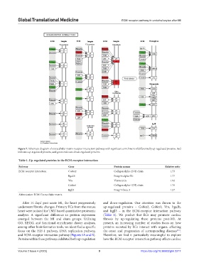
Figure 7. Schematic diagram of extracellular matrix receptor interaction pathway with significant enrichment of differentially up-regulated proteins. Red
indicates up-regulated proteins, and green indicates down-regulated proteins.
Table 1. Up‑regulated proteins in the ECM‑receptor interaction
Pathway Gene Protein names Relative ratio
ECM-receptor interaction Col6α2 Collagen alpha-2(VI) chain 1.73
Itga2b Integrin alpha-IIb 1.77
Vtn Vitronectin 1.92
Col6α1 Collagen alpha-1(VI) chain 1.76
Itgβ3 Integrin beta-3 1.57
Abbreviation: ECM: Extracellular matrix.
After 14 days’ post-acute MI, the heart progressively and down-regulation. Our attention was drawn to the
underwent fibrotic changes. Primary ECs from the mouse up-regulated proteins – Col6α2, Col6α1, Vtn, Itga2b,
heart were isolated for TMT-based quantitative proteomic and Itgβ3 – in the ECM-receptor interaction pathway
analysis. A significant difference in protein expression (Table 1). We predict that ECs may promote cardiac
emerged between the MI and sham groups. Utilizing fibrosis by up-regulating these proteins post-MI. At
GO, KEGG, and functional enrichment cluster analyses, present, an increasing number of studies focus on how
among other bioinformatics tools, we identified a specific proteins secreted by ECs interact with organs, affecting
focus on the HIF-1 pathway, DNA replication pathway, the onset and progression of corresponding diseases .
[13]
and ECM-receptor interaction pathway (Figures 5A and B). Therefore, we find it particularly meaningful to explore
Proteins within these pathways exhibited both up-regulation how the ECM-receptor interaction pathway affects cardiac
Volume 2 Issue 4 (2023) 9 https://doi.org/10.36922/gtm.2217