
Page 99 - GTM-2-4
P. 99
Global Translational Medicine ECM receptor pathway in endotheliocytes after MI
A B
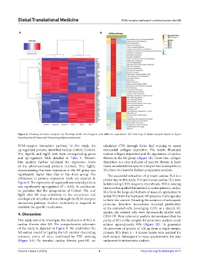
Figure 6. Heatmap of cluster analysis. (A) Heatmap of the first 50 genes with different expressions. (B) Heat map of cluster analysis based on Kyoto
Encyclopedia of Genes and Genomes pathway enrichment.
ECM-receptor interaction pathway. In this study, the calculated CVF through Sirius Red staining to assess
up-regulated proteins identified include Col6α2, Col6α1, myocardial collagen deposition. The results illustrated
Vtn, Itgα2b, and Itgβ3, with their corresponding genes evident collagen deposition and the appearance of cardiac
and up-regulated folds detailed in Table 1. Western fibrosis in the MI group (Figure 1B). Given that collagen
blot analysis further validated the expression levels deposition is a clear indicator of reactive fibrosis in heart
of the aforementioned proteins (Col6α2, Vtn, Itgβ3), tissue, we selected this specific time point to isolate primary
demonstrating that their expression in the MI group was ECs from the heart for further comparative analysis.
significantly higher than that in the sham group. The The successful extraction of primary cardiac ECs is a
differences in protein expression levels are detailed in pivotal step in this study. Primary mouse cardiac ECs were
Figure 8. The expression of target pathway-related proteins isolated using CD31 magnetic microbeads. While existing
was significantly up-regulated (P < 0.05). In conclusion, literature has applied this method to isolate primary cardiac
we postulate that the upregulation of Col6α2, Vtn, and ECs from the lungs and kidneys of mice, its application to
Itgβ3 after MI may contribute to the occurrence and isolate ECs from the heart post-MI presents challenges due
development of cardiac fibrosis through the ECM-receptor to their low content. Ensuring the accuracy of subsequent
interaction pathway. Further verification is required to proteome detection necessitates maximal purification
elucidate the specific mechanism. of the extracted cells. Leveraging CD31 as a specific EC
4. Discussion marker, the isolated cells were fluorescently labeled with
CD31-PE. Flow cytometry analysis demonstrated that the
This study aimed to investigate the mechanism of ECs in purity of ECs in the samples for proteomic analysis could
cardiac fibrosis after MI. The comprehensive schematic achieve approximately 89% (Figure 2B). To guarantee
of the study is depicted in Figure 9. We established the the provision of protein of 100 µg from a single sample,
MI animal model by ligating the left anterior descending primary ECs from 6 – 8 mouse hearts were isolated for
coronary artery of mice, confirmed by TTC staining each sample. Subsequent to mass spectrometry, the data
(Figure 1A). To simulate cardiac fibrosis post-MI, we underwent bioinformatics analysis.
Volume 2 Issue 4 (2023) 8 https://doi.org/10.36922/gtm.2217