
Page 102 - GTM-3-3
P. 102
Global Translational Medicine Rheumatoid nodule versus fibrocaseous tubercle
located 1.5 – 2 cm apart and a third similar hazelnut-sized light-green-orcein and collagen-specific picrosirius
4
lesion. The surrounding lung tissue exhibited a prominent red F3BA staining (Figure 7). Immunohistochemical
5,6
alveolar pattern. Microscopically, an eosinophilic necrotic analysis revealed that CD3 T-lymphocytes dominated
+
area was identified, surrounded by a demarcation zone the inflammatory infiltrate, along with CD20 B-cells and
+
of histiocytes and multinucleated macrophages. Residual CD68 histiocytes (Figure 8). Acid-fast bacilli were not
+
blood vessel structures were present within the necrotic detected using Ziehl-Neelsen staining. However, based on
3
area, confirmed by elastic fiber staining. A medium- the characteristic histology, the lesion was diagnosed as a
sized blood vessel located near the lesion was found RhNod, accompanied by systemic rheumatoid vasculitis,
to have its lumen filled with granulation tissue. The interstitial pneumonitis, and pleuritis. The seropositive
interstitial alveolar septa were widened. Ziehl-Neelsen RA was well controlled with steroid therapy (Medrol).
staining revealed no acid-fast bacilli, and periodic Figures 6-11 illustrate the histological characteristics of the
acid–Schiff staining demonstrated no fungal elements RhNod from the tissue samples submitted for consultation.
(Aspergillus). One of the authors, Miklós Bély, a consultant
pathologist, histologically confirmed the presence of 3. Discussion
a confluent rheumatoid nodule (RhNod) surrounded The risk of TB is higher in patients with RA than in the
by a demarcation zone of fibroblasts, fibrocytes, and general population due to the impaired immune response
histiocytes, including macrophages (Figure 6D-F). Within in elderly patients with autoimmune disease. 7-12 The
the fibrinoid necrotic debris, remnants of blood vessels presence of fibrocaseous tuberculosis further increases the
were demonstrated using combined elastic fiber-specific
risk of endogenous exacerbation and miliary dissemination
(mTB) of dormant TB. The risk of mTB and a fatal outcome
A D
A D
B E
B E
C F
C F
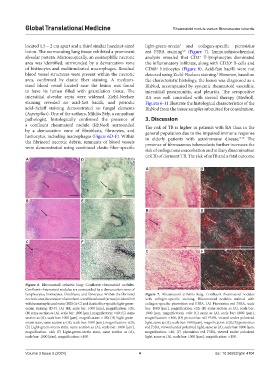
Figure 6. Rheumatoid arthritis lung: Confluent rheumatoid nodules.
Confluent rheumatoid nodules are surrounded by a demarcation zone of
lymphocytes, histiocytes, fibroblasts, and fibrocytes. Within the fibrinoid Figure 7. Rheumatoid arthritis lung: Confluent rheumatoid nodules
necrotic area, the contour of a medium-sized blood vessel (arrows) is identified with collagen-specific staining. Rheumatoid nodules stained with
with hematoxylin and eosin (HE) (A-C) and elastic fiber-specific light-green- collagen-specific picrosirius red F3BA. (A) Picrosirius red F3BA, scale
orcein staining (D-F). (A) HE, scale bar: 1000 [µm], magnification: ×20; bar: 1000 [µm], magnification: ×20; (B) same section as (A), scale bar:
(B) same section as (A), scale bar: 1000 [µm], magnification: ×40; (C) same 1000 [µm, magnification: ×40; (C) same as (A), scale bar: 1000 [µm],
section as (A), scale bar: 1000 [µm], magnification: ×100; (D) Light-green- magnification: ×100; (D) picrosirius red F3BA, viewed under polarized
orcein stain, same section as (A), scale bar: 1000 [µm], magnification: ×20; light, same as (A), scale bar: 1000 [µm], magnification: ×20; (E) picrosirius
(E) Light-green-orcein stain, same section as (A), scale bar: 1000 [µm], red F3BA, viewed under polarized light, same as (A), scale bar: 1000 [µm,
magnification: ×40; (F) Light-green-orcein stain, same section as (A), magnification: ×40; (F) picrosirius red F3BA, viewed under polarized
scale bar: 1000 [µm], magnification: ×100. light, same as (A), scale bar: 1000 [µm], magnification: ×100.
Volume 3 Issue 3 (2024) 4 doi: 10.36922/gtm.4104