
Page 105 - GTM-3-3
P. 105
Global Translational Medicine Rheumatoid nodule versus fibrocaseous tubercle
A B distinction between fibrocaseous tuberculosis and RhNod
crucial in clinical practice. This case series illustrates the
histological differences between these two pathological
entities. The key histological sign for RhNod is the presence
of blood vessel remnants within the fibrinoid necrotic area,
reflecting its vascular origin. The detection of inflamed
blood vessels elsewhere in the lung, such as non-specific
C D fibrinoid necrotic and/or granulomatous autoimmune
vasculitis, along with the possible co-existence of
interstitial pneumonitis (with or without pleuritis), further
supports the rheumatoid nature of the process. In contrast,
tuberculous necrosis is characterized by coalescent
necrosis that does not respect anatomical borders (without
structural remnants of the lung), which is a hallmark of the
tuberculous process.
E F
Acknowledgments
None.
Funding
None.
G H
Conflict of interest
The authors declare that they have no competing interests.
Author contributions
Conceptualization: Miklós Bély
Data curation: Ágnes Apáthy
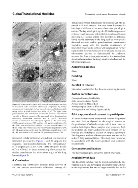
Figure 12. Rheumatoid arthritis with systemic autoimmune vasculitis Formal analysis: Miklós Bély
in association with co-existent tuberculosis complicated by miliary Writing-original draft: Miklós Bély
dissemination. Depicted are the heart, a small intramural (subepicardial) Writing-review and editing: Ágnes Apáthy
artery, and an arteriole (marked by a white ellipse). The figure shows
granulomatous and rheumatoid nodule-like necrotic autoimmune Ethics approval and consent to participate
vasculitis in different segments of the same small artery. Granulomatous
necrotizing autoimmune vasculitis with a typical rheumatoid All case descriptions are anonymized. Neither the patients
nodule is observed in the subsequent segment of the same artery. nor their families objected to the surgical biopsies or
(A) Hematoxylin and eosin (HE), scale bar: 1250 [µm], magnification: ×50; the publication of related excerpts, which were based on
(B-D) HE, same section as (A), scale bar: 1250 [µm], magnification: ×125;
(E and F) HE, same section as (A), scale bar: 100 [µm], magnification: ×200; routine histological tissue samples and protocols. Both
(G) HE, same section as (A), scale bar: 1250 [µm], magnification: ×125; the patients and their families provided consent for the
(H) HE, same section as (G), scale bar: 100 [µm], magnification: ×200. biopsies and histological analysis of surgical specimens.
The diagnostic analysis of these specimens was part of the
interstitial cellular infiltration around foci reminiscent of authors’ daily responsibilities. This study was conducted
interstitial pneumonitis (Figures 1 and 2) supports this in accordance with the local legislation and institution
diagnosis. Immunohistochemically, the predominance requirements.
of T-lymphocytes (CD3, CD4, CD8) alongside B-cells
(CD20, CD79α) in areas surrounding fibrocaseous foci Consent for publication
further corroborates the tuberculous origin of the lung The study subjects gave consent to publish their data.
lesions (Figure 5).
Availability of data
4. Conclusion
The data from our study can be shared anonymously. The
Differentiating tuberculous necrosis from necrosis in original clinical and histological documents were archived
RA can present considerable difficulties, making the in our department, but are no longer available due to the
Volume 3 Issue 3 (2024) 7 doi: 10.36922/gtm.4104