
Page 100 - GTM-3-3
P. 100
Global Translational Medicine Rheumatoid nodule versus fibrocaseous tubercle
clinical histories of two RA patients. The findings are based A B
on a unique autopsy population and biopsy material from
the Department of Pathology, as well as material received
in consultation over nearly 50 years.
2. Case presentation
2.1. Case 1
A 56-year-old female patient with a long-standing history C D
of seropositive RA was being managed effectively with
leflunomide (Sanofi-Aventis, Deutschland), remaining
largely asymptomatic. During a routine lung screening, a
bifocal lung lesion was detected. Chest X-ray and computed
tomography scan, performed at the Pulmonology Clinic
of Semmelweis University, revealed a 15 × 18 mm well-
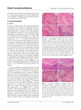
circumscribed lesion in the middle lobe of the right lung. Figure 1. Co-existent seropositive rheumatoid arthritis and fibrocaseous
Furthermore, a 27 × 17 mm lesion with broad pleural tuberculosis. Histopathological findings show erosive coalescent
contact was noted. No further abnormalities were identified fibrocaseous tubercles in the lung of a patient with seropositive
in the lungs or mediastinum, and no enlarged lymph nodes rheumatoid arthritis, along with an adjacent obliterated medium-sized
were detected. The pleural surfaces appeared unaffected. pulmonary artery and an eroded small artery junction (indicated by
arrow). The caseous necrosis does not respect anatomical borders. The
Differential diagnoses include metastatic tumor, abscess, fibrocaseous necrotic center is partially boarded by basophilic debris,
or a specific tuberculous process. Lung surgery, including consisting of granulocytes and lymphocytes (differentiated plasm cells were
intraoperative lung biopsy and wedge resection involving not observed). Interstitial cellular infiltration (reminiscent of interstitial
two segments, was performed. A gross examination of a pneumonitis) is found only close to the tuberculous foci. (A) Hematoxylin
6 × 6 × 4 cm lung segment with a smooth external surface and eosin staining (HE), scale bar: 1000 [µm], magnification: ×20;
(B) same section as (A), HE, scale bar: 1000 [µm], magnification: ×40;
revealed a walnut-sized, lobed abscess containing thick, (C) same section as (A), HE, scale bar: 1000 [µm], magnification: ×100;
yellowish materials. The abscess was partially encapsulated (D) same section as (A), HE, scale bar: 100 [µm], magnification: ×200.
by a dense fibrotic wall. Histological examination suggested
a tuberculous process, while infection agents, such as A B
Echinococcus and Aspergillus, as well as malignancy, were
ruled out. Ziehl-Neelsen staining did not demonstrate the
2
presence of acid-fast bacilli, and repeated cultures were
3
negative.
The patient underwent antituberculosis treatment with
levofloxacin, amoxicillin, clavulanate potassium, and later
clindamycin. However, the treatment was discontinued C D
due to liver dysfunction and in the absence of definitive
evidence of TB. The surgical specimen was reviewed by
Miklós Bély, who confirmed histologically the presence
of a coalescent fibrocaseous tubercle, bordered partly by a
wide zone of granulation tissue with epithelioid cells and
partly by sclerotic fibrous tissues (Figures 1-3). Adjacent
to the tuberculous foci, an obliterated middle-sized artery
with erosion at a small artery junction was identified Figure 2. Co-existent seropositive rheumatoid arthritis and
(Figure 1). Combined staining with elastic fiber-specific fibrocaseous tuberculosis. A fibrocaseous tubercle is surrounded
by granulation tissue composed of epithelioid histiocytes and
light green-orcein and collagen-specific picrosirius Langhans-type multinucleated giant cells (indicated by arrows),
4
red F3BA demonstrated damage to the vascular wall with accompanying lymphoid cell infiltration (differentiated plasma
5,6
(Figure 4). Immunohistochemically, CD3 T-lymphocytes cells were not observed). (A) Hematoxylin and eosin staining (HE),
+
and CD20 B-lymphocytes were observed infiltrating the scale bar: 1000 [µm], magnification: ×40; (B) same section as (A),
+
granulation tissue in the demarcation zone, along with HE, scale bar: 1000 [µm], magnification: ×100; (C) same section as
(A), HE, scale bar: 100 [µm], magnification: ×200; (D) same section
CD68 histiocytes. Notably, differentiated B-cells (plasma as (A) showing Langhans-type giant cells, HE, scale bar: 1000 [µm],
+
cells) were absent (Figure 5). Although acid-fast bacilli magnification: ×200.
Volume 3 Issue 3 (2024) 2 doi: 10.36922/gtm.4104