
Page 104 - GTM-3-3
P. 104
Global Translational Medicine Rheumatoid nodule versus fibrocaseous tubercle
A D derived lymphocytes (T-cells), with a normal proportion
of CD4 (OKT4 ) helper/inducer and CD8 (OKT8 )
+
+
suppressor/cytotoxic subpopulations. 20,22 The histology of
our patients aligns with the descriptions mentioned above.
Furthermore, we observed a moderate T-cell dominance
in the lymphoid mantle of the fibrocaseous tubercle,
alongside B-cells, in contrast to the sparse (scattered)
B E perinodal lymphoid infiltration of the RhNod, with no
differentiated plasma cells detected. The pathogenesis of
RhNods remains contentious. 18,20,21 Factors such as vascular
occlusion, neutrophil involvement, immune complexes,
tissue lesions, and local complement activation due to
pressure and movement are thought to contribute to their
formation.
23
C F According to Ziff, as quoted by Mohr, trauma and
microhemorrhages stimulate monocytes and macrophages,
which release immune complexes and various chemotactic
and toxic agents (e.g., proteinases, collagenases, cytotoxic
substances), resulting in necrosis surrounded by a cellular
demarcation zone. Sokoloff and Bunim emphasized the
24
role of vasculitis in the pathogenesis of RhNods. Koizumi
et al. described RhNods as the most severe form of
25
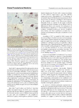
Figure 11. Rheumatoid arthritis lung: Rheumatoid nodule with B-cell autoimmune granulomatous necrotizing vasculitis. Our
infiltration. A rheumatoid nodule surrounded by granulation tissue. CD20 previous studies 26,27 histologically confirmed Koizumi
(A-C) and CD79α (D-F) B-cell infiltration is more concentrated compared et al.’s findings (Figure 12).
25
to the more diffuse CD3 T-cell (Figure 10A-C) and CD43 peripheral T-cell
(Figure 10E and F) infiltration. (A) CD20 monoclonal antibody (N1502, In agreement with Mohr and Fassbender, we consider
20
18
DAKO, Denmark), streptavidin-biotin complex/horseradish peroxidase the histology of RhNods to represent a stage-dependent
reaction, scale bar: 1000 [µm], magnification: ×20; (B) same section as (A),
scale bar: 1000 [µm], magnification: ×40; (C) same section as (a), scale pathological process. In the early stage, fibrinoid necrotic
bar: 1000 [µm], magnification: ×100; (D) CD79α monoclonal antibody debris is surrounded by histiocytes, while in later stages,
(N1628, DAKO, Denmark), streptavidin-biotin complex/horseradish a denser core becomes demarcated by fibroblasts and
peroxidase reaction, scale bar: 1000 [µm], magnification: ×20; (E) same fibrocytes. Evidence for the vascular origin of RhNods
section as (d), scale bar: 1000 [µm], magnification: ×40; (F) same section is provided by the presence of remnants of blood vessels
as (E), scale bar: 1000 [µm], magnification: ×100.
within the necrotic center (Figures 6 and 7).
Fassbender emphasized that differentiating tuberculosis The presence of systemic, non-specific, fibrinoid
18
necrosis from necrosis in RA patients under the microscope necrotic, or granulomatous autoimmune vasculitis in
can present considerable difficulties. A caseous tubercle is other areas of the lung, combined with the potential
characterized by central necrosis impregnated with fibrinous co-existence of interstitial pneumonitis (with or without
exudate, surrounded by a broad zone of epithelioid cells, pleuritis), further supports the rheumatoid nature of the
occasionally accompanied by Langhans-type giant cells and disease and confirms RA as the underlying condition. In
a few lymphocytes. According to Zollinger, the absence our interpretation, inflammation of the capillaries is the
18
19
of differentiated B-cells (plasma cells) is characteristic of quintessence of RA-related interstitial pneumonitis, which
lymphocytic infiltration in TB. The histology of a RhNod is can be regarded as a manifestation of systemic autoimmune
similar, with a fibrinoid necrotic center surrounded by a wide vasculitis involving the capillaries. 28
cellular zone of radially arranged histiocytes (macrophages) In contrast to RhNods, anthracotic pigmentation of
and fibroblasts. 18 fibrotic scars, deposition of calcium salts (calcium carbonate
Both Mohr and Gardner and McClure described and calcium phosphate) in fibrocaseous masses, dominant
21
29
20
similar histological features for both caseous tubercles histiocyte infiltration in the demarcation zone around
and fibrinoid necrotic RhNods. Immunohistochemically, the tubercle, T-cell dominance (without the presence of
the fibrinoid necrotic center of RhNod shows positivity plasma cells), and caseous necrosis that does not respect
for IgG, IgM, and complement. 20,21 The lymphocytic anatomical borders (with or without adjacent obliterative
infiltration is sparse, consisting almost entirely of thymus- vasculitis) all suggest a tuberculous origin. Localized
Volume 3 Issue 3 (2024) 6 doi: 10.36922/gtm.4104