
Page 101 - GTM-3-3
P. 101
Global Translational Medicine Rheumatoid nodule versus fibrocaseous tubercle
A B A B
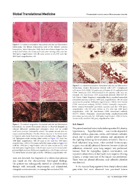
Figure 3. Co-existent seropositive rheumatoid arthritis and fibrocaseous C D
tuberculosis. The fibrous demarcation zone of the tubercle contains
hemosiderin-laden histiocytes, likely due to microhemorrhages from the
eroded small artery. (A) Hematoxylin and eosin staining (HE), scale bar:
1000 [µm], magnification: ×20; (B) same section as (A), HE, scale bar:
1000 [µm], magnification: ×40.
A B
E F
C D
Figure 5. Co-existent seropositive rheumatoid arthritis and fibrocaseous
tuberculosis. Erosive fibrocaseous tubercle with CD3 T-lymphocyte
+
infiltration (A,B), CD20 B-lymphocyte infiltration (C,D), and epithelioid
+
CD68 histiocytes (E,F). Differentiated B-cells (plasma cells) were not
+
detected. (A) Anti-human CD3 monoclonal antibody (RM-9107-R7,
Lab Vision, United Kingdom), streptavidin-biotin complex/horseradish
peroxidase reaction, scale bar: 1000 [µm], magnification: ×40; (B) same
E F section as (A), scale bar: 1000 [µm], magnification: ×100; (C) Anti-human
CD20 monoclonal antibody (N1502, DAKO, Denmark), streptavidin-
biotin complex/horseradish peroxidase reaction, scale bar: 1000 [µm],
magnification: ×40; (D) same section as (C), scale bar: 1000 [µm],
magnification: ×100; (E) Anti-human CD68 monoclonal antibody
(N1577, DAKO, Denmark), streptavidin-biotin complex/horseradish
peroxidase reaction, scale bar: 1000 [µm], magnification: ×100; (F) Same
section as (E), scale bar: 100 [µm], magnification: ×200.
Figure 4. Co-existent seropositive rheumatoid arthritis and fibrocaseous 2.2. Case 2
tuberculosis. An erosive fibrocaseous tubercle is observed alongside an
adjacent obliterated medium-sized pulmonary artery and an eroded The patient’s medical history included seropositive RA, obesity,
small artery junction (indicated by arrow). The caseous necrosis does not hypertension, hypothyroidism, non-insulin-dependent
respect anatomical borders. The fine fibrous structure of the blood vessels diabetes mellitus, glaucoma, stroke, and transient ischemic
is impaired, with elastic fibers being more vulnerable than collagen fibers. attack due to carotid artery stenosis, and amputation of
(A) Light-green-orcein staining (same section as Figure 1C), scale bar: the right leg following femoral artery embolism. Due to a
1000 [µm], magnification: ×40; (B) same as (A), scale bar: 1000 [µm],
magnification: ×100; (C) Picrosirius red F3BA (same section as Figure 1C), focal subpleural lung lesion, video-assisted thoracoscopic
scale bar: 1000 [µm], magnification: ×40; (D) same section as (C), scale surgery was initially planned; however, because of pleural
bar: 1000 [µm], magnification: ×100; (E) same section as (C) viewed under adhesions, extended open lung surgery was performed
polarized light, scale bar: 1000 [µm], magnification: ×40; (F) same section as instead. Tests for Aspergillus, sputum examination, and
(C) viewed under polarized light, scale bar: 1000 [µm], magnification: ×40.
repeated cultures for acid-fast bacilli were negative. During
surgery, a wedge resection of the lingula was performed.
were not detected, the diagnosis of a tuberculous process
was based on the characteristic histological findings. There were no pleural effusions; only adhesive pleuritis
The patient was subsequently started on antituberculous was detected.
therapy with isoniazid, streptomycin, and ethambutol, In the resected lung segment, three sharply demarcated
alongside corticosteroids, to manage her activated RA. gray-white foci were observed: two pea-sized lesions
Volume 3 Issue 3 (2024) 3 doi: 10.36922/gtm.4104