
Page 93 - GTM-3-3
P. 93
Global Translational Medicine SLE patient underwent PD
pancreatic cancers, and lung cancers, with a 3- to 4-fold and low levels of C3 and C4 complement proteins. In
increase in risk. A meta-analysis also demonstrated 2018, due to progressively worsening renal function, he
2,3
that SLE patients have an increased risk of pancreatic underwent renal transplantation. Post-transplant, he was
cancer (hazard ratio = 1.42, 95% confidence interval = maintained on triple immunosuppressive therapy with
1.32 – 1.53). Furthermore, 10 – 20% of SLE patients may tacrolimus, mycophenolate mofetil, and low-dose daily
4
progress to end-stage renal disease, necessitating renal prednisone. Over the past year, his immunosuppressive
transplantation. In addition, several studies suggest that regimen was reduced to tacrolimus (2 mg BID) and
5
cancer occurrence is related to renal transplantation. mycophenolate mofetil (750 mg BID). His last tacrolimus
6
Therefore, SLE patients who have undergone renal blood concentration, measured in September 2022, was
transplantation have an increased incidence of cancer. This within the therapeutic range at 5.75 ng/mL. The serum
increased risk is attributed to a range of factors, the most creatinine level was 71 μmol/L at admission. Given
important of which may be the adverse effects of long-term the patient’s presentation with obstructive jaundice
immunosuppressive medications. These medications, (serum total bilirubin level = 350 μmol/L), percutaneous
3
such as azathioprine and cyclosporin, can impair DNA transhepatic cholecystostomy was performed on the 1 day
st
repair and activate oncogenic pathways. 7 of admission.
Pancreatic cancer is ranked as one of the most lethal The patient underwent PD combined with resection and
and aggressive cancers, with a 5-year survival rate of only reconstruction of the superior mesenteric vein in December
10% in 2020. The poor prognosis is attributed to its rapid 2022. Immunosuppressive drugs were discontinued on
8
progression, early metastasis, and the lack of obvious the day of the procedure (Figure 2). Given the patient’s
clinical symptoms or sensitive screening approaches immunocompromised state, piperacillin–tazobactam
for early-stage detection. Previous studies have shown was administered as a post-operative anti-infective
that SLE is closely associated with an increased risk of prophylaxis regimen for 6 days, along with human gamma
pancreatic cancer. Radical resection remains the most globulin (10 g IV, QD) for 5 days. Immunosuppressive
4
effective treatment option for pancreatic cancer. However,
due to the complexity of pancreaticoduodenectomy (PD),
there is controversy over whether PD is safe for SLE
patients who have undergone renal transplantation. This
case study describes a PD performed for pancreatic cancer
in a 44-year-old male with SLE and a history of renal
transplantation. Our case demonstrates the feasibility of
performing a PD in SLE patients who have undergone
renal transplantation and, for the 1 time, reveals the
st
changes in the immune microenvironment through
in silico analysis.
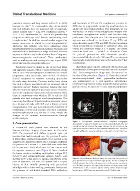
Figure 1. Pre-operative contrast-enhanced computed tomography
2. Case presentation demonstrates a large, low-density mass in the pancreatic head. The red
A 44-year-old male patient was admitted to our triangle on the left indicates the tumor, whereas the red triangle on the
right shows the tumor’s invasion of the superior mesenteric vein. The yellow
Pancreaticobiliary Surgery Department in December triangle highlights the transplanted kidney, implanted in the right iliac fossa.
2022. He presented with diffuse epigastric pain and
jaundice that had developed over the previous 2 weeks.
A triple-phase computer tomography (CT) scan led to the
diagnosis of a pancreatic tumor. The CT images confirmed
the presence of a 38 mm × 27 mm solid mass at the level
of the pancreatic head, which was in close contact with
the superior mesenteric vein (Figure 1). Serum levels of
the tumor markers, including carbohydrate antigen 19-9
(CA19-9) and carcinoembryonic antigen (CEA), were
elevated at 99 U/mL and 6.71 ng/mL, respectively, at the Figure 2. Intra-abdominal findings during the operation (left panel) and
time of admission. Of note, the patient had a 15-year gross specimen after surgical resection (right panel). The black triangle on
the left indicates the end-to-end anastomosis of the superior mesenteric
history of SLE, initially presenting with generalized vein. The black triangle on the right shows the tumor in the pancreatic head.
arthralgia without joint swelling, proteinuria, leukopenia, Abbreviations: CHA: Common hepatic artery; rRHA: Replaced right
the presence of anti-double-stranded DNA antibodies, hepatic artery; SMV: Superior mesenteric vein; SV: Splenic vein.
Volume 3 Issue 3 (2024) 2 doi: 10.36922/gtm.2893