
Page 88 - GTM-3-3
P. 88
Global Translational Medicine rh-PDGF for peri-intrabony defects
This study presents 14 consecutively treated cases options with the patients, and written informed consent
to assess the efficacy of non-surgical management for was obtained.
intrabony defects in the anterior esthetic area, employing Clinical parameters such as PD, gingival recession
a combination of rhPDGF-BB and a collagen sponge. depth, and radiographic bone fill were recorded before and
This minimally invasive approach is designed to achieve 6 months after the surgical intervention (Figures 1-3).
periodontal repair without compromising esthetic
outcomes. Patients received oral hygiene instructions, with an
emphasis on maintaining oral health post-non-surgical
2. Methods periodontal therapy. They were also provided with
7,8
Fourteen systemically healthy, non-smoker, non-diabetic guidance and motivation for effective home care to ensure
subjects with no history of chemotherapy or radiotherapy proper oral hygiene.
were included in this study, conducted at a private practice The treatment began with local anesthesia using 2%
in California, United States of America (USA). These lidocaine and 1:100,000 epinephrine. Ultrasonic scaling,
individuals presented with single intrabony defects in the combined with Gracey curettes, was performed to carefully
anterior region of the maxilla, and all treatments were target the depth of the bony defect, inducing bone scoring
standardized using the same protocol. Before initiating and promoting bleeding. Subsequently, a collagen sponge
treatment, the surgeon thoroughly discussed all treatment soaked with rh-PDGF-BB (GEM21S, Geistlich North
America, USA) was then placed in the intrabony pocket
A B and packed as apically as possible. The collagen sponge was
secured with a vertical mattress suture.
A B
C D
C D
E F
E F
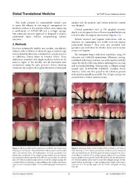
Figure 1. Infrabony defect in the mesial area (tooth #9). (A) Clinical view Figure 2. Infrabony defect noted in distal of #9 in Patient 2. (A) Clinical
showing a 6-mm buccal. The periodontal intrabony pocket was recorded view showing a 7-mm buccal periodontal intrabony pocket measured
with a periodontal probe. (B) Radiographic showing an angular bone with a periodontal probe. (B) Radiograph showing an angular bone defect
defect in the mesial area. (C) A collagen sponge soaked with recombinant in the distal of tooth #9. (C) A collagen sponge soaked with recombinant
human platelet-derived growth factor-BB was inserted into the affected human platelet-derived growth factor-BB inserted into the affected area.
area. (D) Vertical mattress suture used to secure the collagen sponge. (E) (D) Vertical mattress suture used to secure the collagen sponge. (E) Six-
6-month follow-up showing a 2 mm probing pocket depth, indicating a month follow-up showing a 3 mm probing pocket depth, indicating a
reduction of 4 mm pocket depth. (F) 6-month radiograph showing bone reduction of 4 mm pocket depth. (F) Six-month radiograph showing
fill. bone fill.
Volume 3 Issue 3 (2024) 2 doi: 10.36922/gtm.3313