
Page 117 - GTM-4-1
P. 117
Global Translational Medicine AA amyloidosis in rheumatoid arthritis
A B
C D
E F
H G
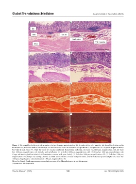
Figure 1. Rheumatoid arthritis, systemic secondary AA amyloidosis, gastrointestinal AA deposits with erosive gastritis. AA deposition is observed in
the submucosa, within the walls of arterioles (a) and small arteries, as well as interstitial collagen fibers (I). Only minimal AA deposits are present within
the walls of small veins (V), while the venule (v) remains unaffected. Hematoxylin and eosin: (A) Scale bar: 1000 µm, magnification: ×20; (B) Scale
bar: 1000 µm, magnification: ×40. Periodic acid–Schiff stain: (C) Scale bar: 1000 µm, magnification: ×20; (D) Scale bar: 1000 µm, magnification: ×40;
Congo red staining (without alcoholic differentiation, covered with gum Arabic): (E) Scale bar: 1000 µm, magnification: ×20; (F) Scale bar: 1000 µm,
magnification: ×40; Congo red staining (without alcoholic differentiation, covered with gum Arabic, and viewed under polarized light): (G) Scale bar:
1000 µm, magnification: ×20; (H) Scale bar: 1000 µm, magnification: ×40.
Notes: fm: Gastric fundic mucosa; mm: muscularis mucosae; Myo: Muscularis propria; sm: Submucosa.
Abbreviation: AA: Amyloid A.
Volume 4 Issue 1 (2025) 109 doi: 10.36922/gtm.5325