
Page 118 - GTM-4-1
P. 118
Global Translational Medicine AA amyloidosis in rheumatoid arthritis
A B
C D
E F
G H
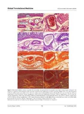
Figure 2. Rheumatoid arthritis, systemic secondary AA amyloidosis, and gastrointestinal AA deposits (same tissue sections shown in Figure 1). AA
deposition is observed in the submucosa, within the walls of arterioles and small arteries, as well as interstitial collagen fibers. Only minimal deposits
within the walls of small veins (yellow arrow), while the venules remain unaffected (black arrow). Hematoxylin and eosin: (A) Scale bar: 1000 µm,
magnification: ×100; (B) Scale bar: 100 µm, magnification: ×200; Periodic acid–Schiff stain: (C) Scale bar: 1000 µm, magnification: ×100; (D) Scale bar:
100 µm, magnification: ×200; Congo red staining (without alcoholic differentiation, covered with gum Arabic): (E) Scale bar: 1000 µm, magnification:
×100; (F) Scale bar: 100 µm, magnification: ×200; Congo red staining (without alcoholic differentiation, covered with gum Arabic, and viewed under
polarized light): (G) Scale bar: 1000 µm, magnification: ×100; (H) Scale bar: 100 µm, magnification: ×200. 52
Abbreviations: a: Arteriole; A: Small artery; mm: Muscularis mucosae; Venule; V: Small vein; AA: Amyloid A.
Volume 4 Issue 1 (2025) 110 doi: 10.36922/gtm.5325