
Page 119 - GTM-4-1
P. 119
Global Translational Medicine AA amyloidosis in rheumatoid arthritis
A B A B
C D
Figure 3. Immunohistochemical Staining of AA deposits using anti-
human AA component. Anti-human AA component, dilution 1:100
(monoclonal antibody MO759, DAKO, Denmark], streptavidin-biotin- E F
complex/horseradish peroxidase reaction: (A) scale bar: 100 µm,
magnification: ×200; (B) scale bar: 100 µm, magnification: ×400.
Abbreviation: AA: Amyloid A.
G H
parallel trend. Organs frequently affected by amyloid
deposition exhibited extensive accumulation, whereas
less frequently involved organs showed comparatively
lower deposit levels. However, an inverse relationship
was observed in the pancreas and GI tract (Table 3 and I J
Figure 6). Figure 6 summarizes the quantitative differences
in prevalence and amount of AA deposits across various
organs in these 34 RA patients.
AA deposition in the kidneys, heart, pancreas, GI tract, K L
liver, lungs, skin, and brain correlated with the average
severity of sAAa. Figure 7 illustrates the distribution of AA
deposition across these organs.
M N
3.6. Characteristics of giAAa
The accumulation of AA deposits in the GI tract followed
a progressive and generally linear trajectory, with an
exponential increase in the terminal stage.
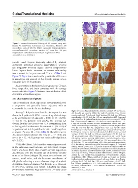
Figure 4. Cecum, rheumatoid arthritis, systemic secondary AA amyloidosis.
Among 31 RA patients with sAAa, AA deposition was AA deposits are observed within the walls of arterioles, while venules
absent in 2 patients (6.45%), representing a latent stage remain unaffected. Periodic acid–Schiff staining: (A) Scale bar: 1250 µm,
of GI amyloidosis (AA deposits = 0.00). In 17 (54.84%) magnification: ×50; (B) Scale bar: 125 µm, magnification ×125. Congo red
staining (without alcoholic differentiation, covered with gum Arabic): (C) Scale
of the 31 RA patients with giAAa, the average AA bar: 1250 µm, magnification: ×50; (D) Scale bar: 125 µm, magnification ×125.
deposit level in the GI tract was <0.8, categorizing them Congo red staining (without alcoholic differentiation, covered with gum
as having mild involvement. In contrast, 12 (38.71%) of Arabic, viewed under polarized light): (E) Scale bar: 1250 µm, magnification:
×50; (F) Scale bar: 125 µm, magnification ×125. AA deposits exhibit
31 patients had AA deposit levels >0.8, classifying them birefringence under polarized light, appearing as characteristic apple-green
as severe (Patients 20 – 31 in Table 4). The difference in birefringence. Congo red staining (without alcoholic differentiation, covered
AA deposit levels between the mild (n = 17, <0.8) and with gum Arabic): (G) Scale bar: 1250 µm, magnification: ×50; (H) Scale bar:
125 µm, magnification ×125. Performate pretreatment for 1 s and Congo
severe (n = 12, >0.8) groups was statistically significant red staining (without alcoholic differentiation, covered with gum Arabic,
(P < 0.000). viewed under polarized light): (I) Scale bar: 1250 µm, magnification: ×50;
(J) Scale bar: 125 µm, magnification ×125. After performate pretreatment
Within the GI tract, AA deposition was most pronounced (1 s), the staining intensity (congophilia) increases compared to Congo red
in the arterioles, small arteries, and interstitial collagen staining without pretreatment. AA protein deposits are highly sensitive to
fibers, which are likely sites of early amyloid deposition. performate pretreatment; birefringence disappears immediately, regardless
of amyloid deposition quantity or disease stage. KMnO oxidation (10 min)
4
AA deposits were less frequent and of moderate severity and Congo red staining (without alcoholic differentiation, covered with gum
in reticulin fibers (collagen III), medium-sized veins and Arabic): (K) Scale bar: 1250 µm, magnification: ×50; (L) Scale bar: 125 µm,
magnification ×125. KMnO oxidation (10 min) and Congo red staining
arteries, small veins, and the basement membranes of (without alcoholic differentiation, covered with gum Arabic, viewed under
4
GI glands, reflecting a more advanced stage of amyloid polarized light): (M) Scale bar: 1250 µm, magnification: ×50; (N) Scale
deposition. Rare and minimal AA deposits were observed bar: 125 µm, magnification ×125. AA protein deposits exhibit resistance
to KMnO oxidation (30 s – 1 min), resistance/sensitivity (3 – 5 min), and
in myocytes, venules, and nerves, indicating late-stage, sensitivity (10 min).
4
pre-mortem (terminal) stage amyloidosis (see data under Abbreviation: AA: Amyloid A.
Volume 4 Issue 1 (2025) 111 doi: 10.36922/gtm.5325