
Page 122 - GTM-4-1
P. 122
Global Translational Medicine AA amyloidosis in rheumatoid arthritis
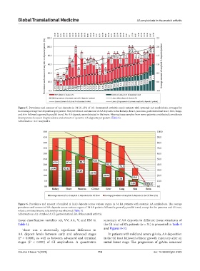
Figure 5. Prevalence and amount of AA deposits in 34 (21.12% of 161 rheumatoid arthritis cases) patients with systemic AA amyloidosis, arranged by
increasing average AA deposition per patient. The prevalence and amount of AA deposits in the kidneys, heart, pancreas, gastrointestinal tract, liver, lungs,
and skin followed a generally parallel trend. No AA deposits were detected in the brain. Missing tissue samples from some patients contributed to moderate
discrepancies between the prevalence and amount of systemic AA deposits per patient (Table 3).
Abbreviation: AA: Amyloid A.
Figure 6. Prevalence and amount of amyloid A (AA) deposits across various organs in 34 RA patients with systemic AA amyloidosis. The average
prevalence and amount of AA deposits across various organs of 34 RA patients followed a generally parallel trend, except for the pancreas and GI tract,
where a minimal inverse relationship was observed (Table 3).
Abbreviations: AA: Amyloid A; GI: gastrointestinal; RA: Rheumatoid arthritis.
tissue classification variables ret, VV, AA, V, and BM in summary of AA deposits in different tissue structures of
Table 4). the GI tract of RA patients (n = 31) is presented in Table 4
There was a statistically significant difference in and Figures 8-10.
AA deposit levels between early and advanced stages In patients with mild and severe giAAa, AA deposition
(P < 0.000), as well as between advanced and terminal in the GI tract followed a linear growth trajectory after an
stages (P < 0.000) of GI amyloidosis. A quantitative initial latent stage. The progression of giAAa remained
Volume 4 Issue 1 (2025) 114 doi: 10.36922/gtm.5325