
Page 123 - GTM-4-1
P. 123
Global Translational Medicine AA amyloidosis in rheumatoid arthritis
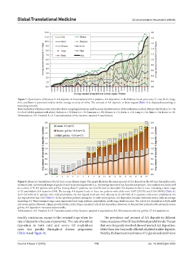
Figure 7. Quantitative differences in AA deposits in rheumatoid arthritis patients. AA deposition in the kidneys, heart, pancreas, GI tract, liver, lungs,
skin, and brain is presented relative to the average severity of sAAa. The amount of AA deposits in these organs (Table 3) is displayed according to
increasing severity.
Note: Individual variations were primarily due to sampling limitations and the semi-objective nature of the evaluation method. Patient distribution (n = 34,
21.1% of 161 RA patients with sAAa): Kidney: n = 33; Heart: n = 33; Pancreas: n = 29; GI tract: n = 31; Liver: n = 32; Lung: n = 34; Skin: n = 24; Brain: n = 26.
Abbreviations: AA: Amyloid A; GI: Gastrointestinal; sAAa: Systemic amyloid A amyloidosis.
Figure 8. Mean AA deposition in the GI tract across disease stages. The graph illustrates the mean amount of AA deposits in the GI tract during the early,
advanced, late, and terminal stages of giAAa, based on increasing severity (i.e., the average amount of AA deposits per patient). This analysis was conducted
in a cohort of 31 RA patients with giAAa. Among these 31 patients, two (6.45%) had no detectable AA deposits in the GI tract, indicating a latent stage
of GI amyloidosis (AA deposits=0.00). The average AA deposit levels in these two patients with sAAa were 1.097 (237/70) and 0.524 (90/85) (Table 4).
In 17 (54.84%) of 31 patients with mild amyloidosis, the AA deposit level was <0.8, whereas in 12 (38.71%) of 31 patients with severe amyloidosis, the
AA deposit level was >0.8 (Table 4). In the past four patients among the 12 with severe giAAa, AA deposition became extremely severe, with an average
exceeding 1.4. These terminal-stage cases represented end-stage systemic amyloidosis, predicting a fatal outcome. The rate of AA deposition in both mild
and severe giAAa followed a linear growth pattern, indicating a consistent rate of AA deposition. However, in the past four patients with extremely severe
giAAa, AA deposition increased exponentially.
Abbreviations: AA: Amyloid A; GI: Gastrointestinal; sAAa: Systemic amyloid A amyloidosis; RA: Rheumatoid arthritis; giAAa: GI AA amyloidosis.
steadily continuous, except in the terminal stage when the The prevalence and amount of AA deposits in different
rate of deposition became exponential. The rate of amyloid tissue structures of the GI tract followed parallel trends. Tissues
deposition in both mild and severe GI amyloidosis that were frequently involved showed marked AA deposition,
cases was parallel throughout disease progression while those less frequently affected exhibited milder deposits.
(Table 4 and Figure 8). Notably, the basement membrane of GI glands and small veins
Volume 4 Issue 1 (2025) 115 doi: 10.36922/gtm.5325