
Page 81 - GTM-4-1
P. 81
Global Translational Medicine Gelatin-based cell carriers for tooth-germ organoids
3.1.2. Methacrylate gelatin microparticles storage. The gentle processing conditions allow for the
Photocrosslinking offers a simple and reproducible integration of proteins, drug compounds, or cells during the
26,27
approach for preparing GelMA HG. To increase the surface fabrication of HMPs. Microsphere sizes were selected by
24
passing them through cell strainers with the desired pore
area, the crosslinked GelMA HG was frozen at −80°C or sizes (Figure 1D). Preparation of Gel MS typically takes
18
in liquid nitrogen, then micronized using a mortar and approximately 8 h, including glutaraldehyde neutralization
pestle (Figure 1B). Mechanical fragmentation is a widely and multiple washes. The average size of the Gel MS used
adopted technique due to its simplicity and efficiency in in this study was 109.47 ± 19.15 µm (Figure 2C and G).
producing hydrogel microparticles (HMPs). This method Previous studies have shown that this size is most efficient
facilitates the rapid generation of substantial quantities of for supporting cell growth, as opposed to larger sizes of Gel
25
MP in a single, streamlined procedure. In this study, the MS. Once lyophilized, Gel MS remains stable at 4°C and
18
process yielded MP with a heterogeneous range of sizes and is suitable for off-the-shelf use.
shapes, completed in approximately 1 h. The average size
(measured along the longitude axis) was 147.83 ± 54.02 µm As shown in Figure 2, GelMA MS and Gel MS displayed
(Figure 2B and F). Lyophilized GelMA MP remains stable relatively uniform spherical shapes, while GelMA MP
when stored at 4°C and can be used off-the-shelf. exhibited a range of sizes and shapes. In addition, the
surfaces of the GelMS, GelMA MP, and GelMA HG
3.1.3. Methacrylate gelatin hydrogels appeared smooth (Figure 2), while the surface of the GelMA
MS showed notable surface topography, likely resulting
Encapsulating cells in photocrosslinked GelMA HG is a from the harsh processing conditions at high temperatures
commonly used method for cell delivery due to its ease and extended washing steps. The key characteristics of
of size and shape determination and the low cytotoxicity each cell carrier are summarized in Table 1.
of photocrossslinking on cells. 21,22 In this study, hDPSCs
were mixed with a solubilized GelMA solution and 3.2. Evaluation of hDPSC growth on/in various cell
encapsulated within the GelMA HG matrix using LAP- carriers
initiated photocrosslinking (Figure 1C). The encapsulation To assess the ability of different carriers to support
process took approximately 30 min post-cell preparation. hDPSC growth, GelMA MS, GelMA MP, and Gel MS
GelMA HG without cells appeared transparent and smooth were rehydrated using equal dry weights, while GelMA
(Figure 2D and H). The GelMA HG with embedded cells HG was prepared with an equivalent weight of GelMA
demonstrated a uniform distribution throughout both powder. Equal numbers of hDPSCs were then seeded onto
the surface and interior of the construct, as expected. HG each carrier or encapsulated within GelMA HG. After
dimensions can be easily adjusted by modifying the mold 10 days of culture, cell-laden GelMA MS, GelMA MP, and
and gel volume; in this study, a 6 mm diameter and 1 mm Gel MS carriers formed aggregates (Figure 3A[v–vii]),
thickness were used. Encapsulated hDPSCs in GelMA whereas the corresponding cell-free controls remained
HG served as the reference for comparison with other cell separate (Figure 3A[ix–xi]), consistent with previous
carriers. studies conducted using only GelMA MS. Viable
7
cells on each carrier were visualized by calcein AM
3.1.4. Gelatin microspheres
staining, where green fluorescence indicated cell viability
In this configuration, gelatin (rather than GelMA) (Figure 3A). Fluorescent intensity was strongest on Gel MS
was used as the starting material. Gel MS were formed (Figure 3A[iii]) and particularly prominent on the surfaces
using the water-in-oil emulsion technique, followed by of GelMA MS and GelMA MP (Figure 3A[i and ii]). In
glutaraldehyde-mediated crosslinking and subsequent GelMA HG, higher cell viability was observed on the HG
washing, filtration, and lyophilization for long-term surface than within the matrix (Figure 3A[iv]).
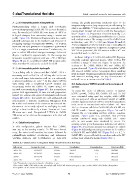
Table 1. Key characteristics of the gelatin-based cell carriers used in this study
Cell carrier Average size (μm) Shapes Crosslinking Processing time Batch variability
GelMA MS 134.8±69.2 Spheres Thermal 2 days Medium
GelMA MP 147.8±54.0 Irregular Photo 1 h Minimal
Gel MS 109.5±19.1 Spheres Glutaraldehyde 8 h Minimal
GelMA HG 6×1 a Disc Photo 0.5 h Minimal
a
Note: Average size is reported in diameter × thickness in mm.
Abbreviations: GelMA HG: Methacrylate gelatin hydrogel; GelMA MP: Methacrylate gelatin microparticles; GelMA MS: Methacrylate gelatin
microspheres; Gel MS: Gelatin microspheres.
Volume 4 Issue 1 (2025) 73 doi: 10.36922/gtm.5897