
Page 82 - GTM-4-1
P. 82
Global Translational Medicine Gelatin-based cell carriers for tooth-germ organoids
A
B
C
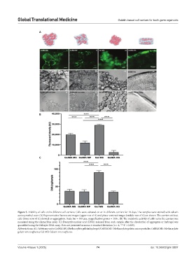
Figure 3. Viability of cells on/in different cell carriers. Cells were cultured on or in different carriers for 10 days. The samples were stained with calcein
acetoxymethyl ester. (A) Representative fluorescent images (upper row of A) and phase-contrast images (middle row of A) are shown. The carriers without
cells (lower row of A) showed no aggregation. Scale bar = 100 µm; magnification power = 100×. (B) The metabolic activity of cells on/in the carriers was
measured using the alamarBlue assay. (C) Deoxyribonucleic acid (DNA) released from each sample after the dissolution of aggregates or hydrogel was
quantified using the Helixyte DNA assay. Data are presented as mean ± standard deviation (n = 4; ***P < 0.005).
Abbreviations: AU: Arbitrary units; GelMA HG: Methacrylate gelatin hydrogel; GelMA MP: Methacrylate gelatin microparticles; GelMA MS: Methacrylate
gelatin microspheres; Gel MS: Gelatin microspheres.
Volume 4 Issue 1 (2025) 74 doi: 10.36922/gtm.5897