
Page 95 - GTM-4-1
P. 95
Global Translational Medicine Brain morphology in obesity
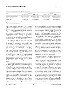
Table 1. Porsolt test indices of the experimental animals
Index Male rats Female rats
Control (n=13) HCD (n=14) Control (n=14) HCD (n=13)
Time of the first swimming act (s) 258.00 (203.00, 320.00) 137.50 (85.00, 240.00)* 260.00 (226.00, 340.00) 265.00 (170.00, 360.00)
Number of freezing 6.00 (3.00, 11.00) 13.00 (9.00, 18.00)* 3.50 (2.00, 7.00) 8.00 (0.00, 13.00)
Freezing time (s) 10.00 (4.00, 13.00) 34.50 (13.00, 61.00)** 3.50 (2.00, 13.00) 8.00 (0.00, 21.00)
Notes: Data are presented as median (25 percentile; 75 percentile); statistically significant differences between the male HCD and male control
th
th
groups at *P<0.05, **P<0.01.
Abbreviation: HCD: High-calorie diet.
system metabolites, such as decreased levels of glutamate date, molecular mechanisms linking obesity, diabetes, and
and choline-containing compounds in the hippocampus. neurodegenerative diseases have been actively investigated,
However, these effects were not associated with markers and a link has been demonstrated in many studies. Our
of central inflammation. The study by Guillemot-Legris et data provide direct evidence for the development of
al. in mice fed HCD showed that inflammatory processes neurodegenerative processes in the PFC and hippocampus
20
were differentially manifested in different brain regions, of rats on a HCD. However, glial activation in the brain
potentially associated with astrocytes, but not microglial areas studied does not always accompany visceral obesity.
cells. In addition, evidence suggests that a HCD potentiates In experiments on male mice, metabolic disturbances
the formation of amyloid plaques in the brains of male and caused by long-term consumption of a high-fat diet were
female APP/E4 mice, but not in APP/E3 mice. 21 shown to increase neuronal oxidative stress and insulin
The analysis of literature data shows that many resistance through suppression of the adiponectin receptor
questions related to the mechanisms of obesity influence (AdipoR1). The authors showed that a high-fat diet
23
on metabolic and morpho-functional characteristics of provokes microglial activation and neuroinflammation in
neurons remain controversial. In our studies (using the the cortical and hippocampal regions of mice. However,
Nissl staining method), pronounced glial activation was suppression of AdipoR1 increased the amyloidogenic
observed in the PFC of male rats with visceral obesity. pathway both in vivo and in vitro. In summary, the authors
However, this phenomenon was not observed in female concluded that excessive fat consumption has a significant
rats, though significant neurodegenerative changes impact on brain function, including an increase in cognitive
were found in their PFC. Our study of hippocampal impairment due to increased oxidative stress associated
morphology showed a variety of changes in different areas with obesity, insulin resistance, neuroinflammation, and
of this brain region, In males, signs of neurodegeneration suppression of AdipoR1 signaling in the brain. 23
were observed in all areas examined (CA1 – CA4), with Our studies show that a HCD leads to depression-like
glial activation occurring only in the CA2 area. In females, behavior in male rats under conditions of “unavoidable
neurodegenerative changes were more selective, involving stress” but has no significant effect on the behavioral
the CA1, CA2, and CA4 areas, while glial activation was strategy of female rats under the same conditions. Several
absent. published studies have noted that peripheral metabolic
The literature generally suggests that chronic inflammation abnormalities in obesity are associated with different
of adipose tissue may induce neuroinflammation and clinical health outcomes and may have sex differences.
hippocampal dysfunction, contributing to the development In one study, a long-term high-fat diet for 16 weeks
24
of cognitive deficits. However, the search for mechanisms was shown to impair glucose metabolism in the brain.
10
of the effect of obesity on hippocampal functional activity Behavioral tests of spatial memory, including the Morris
leads to equivocal findings. For example, it has been shown water maze and Y-maze, showed that memory performance
that high-density leptin receptors are expressed in several was impaired only in male rats on the high-fat diet. In
brain regions involved in higher cognitive functions, addition, a significant decrease in glucose metabolism
including the hippocampus. In addition, leptin has a was observed in male rats, but not in female rats, on this
cognitive stimulatory effect in the hippocampus, and leptin diet. Analysis of genes related to glucose metabolism and
deficiency or insensitivity to leptin results in significant Alzheimer’s disease (AD) pathology in the hippocampus
memory deficits. In the context of obesity, several showed that the expression of glucose transporter 3
22
mechanisms of leptin resistance have been discussed, which (GLUT3), insulin receptor substrate 2 (IRS2), and insulin-
may increase the risk of neurodegenerative diseases. To degrading enzyme (IDE) was significantly reduced in male
15
Volume 4 Issue 1 (2025) 87 doi: 10.36922/gtm.5000