
Page 47 - GTM-4-3
P. 47
Global Translational Medicine Evolution of tunneling techniques
Raetzke observed a mean gain in keratinized gingiva of However, the tunnel procedure resulted in statistically
3.54 mm and a mean residual recession depth of 0.67 mm. greater gains in keratinized tissue width as well as less post-
Notably, the benefits of the technique that the author operative morbidity and pain.
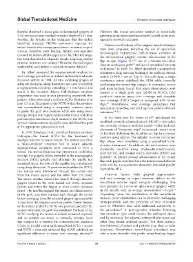
identified included PROMs commonly reported for Various modifications of the original tunnel technique
tunnel-based root coverage procedures—minimal surgical have been proposed, including the use of specialized
trauma, favorable early healing, limited post-operative microsurgical instruments, full-thickness elevation of
7
discomfort, and an esthetic appearance. Raetzke’s technique the interproximal gingiva, coronal advancement of the
9
has been described as elegantly simple, requiring neither flap margin (Figure 2), 8,9,23 use of a biomaterial rather
external incisions nor sutures. However, the technique’s than an autologous graft, and use of specialized suturing
5
8,9
applicability was limited to isolated recession defects. 4
techniques. In 2010, Dr. Allen described a subpapillary
8
8
Dr. Allen presented the supraperiosteal envelope for continuous sling suturing technique for acellular dermal
5
root coverage procedures at isolated and multiple adjacent matrix (ADM) + tunnel flap. In this technique, a single
recession defects in 1994. At sites exhibiting gingiva of continuous suture stabilized the ADM while coronally
adequate thickness, sharp dissection was used to establish positioning the tunnel flap margin. A systematic review
a supraperiostal envelope extending 3–5 mm lateral and and meta-analysis found that when observations were
apical to the recession defects. Full-thickness envelope limited to a single graft type (ADM or SCTG), CAF
preparation was used at sites presenting excessively thin produced superior mean root coverage and complete
gingiva. Allen advocated a uniform SCTG thickness of at root coverage (CRC) frequency compared with tunnel
least 1.5 mm. Placement of the SCTG within the envelope flaps. Nevertheless, root coverage procedures that
23
was accomplished using a temporary mattress suture incorporate tunneling may yield superior patient-oriented
to guide the graft into position while also using tissue outcomes. 7-9,22,23
forceps. Simple interrupted sutures at the mesial and distal 24
graft margins introduced slight tension in the SCTG, and In the same year, Dr. Aroca et al. introduced the
vertical mattress sutures in papilla areas stabilized the graft modified coronally advanced tunnel (MCAT)—also called
at the appropriate apicocoronal level. the coronally advanced modified tunnel—which included
placement of “composite stops” at proximal contact areas
In 1999, Zabalegui et al. modified Raetzke’s envelope to facilitate stabilizing the facial/buccal flap in a coronal
6
technique—the tunnel SCTG—for the treatment of position using sutures. The sulcular incisions and tunnel
multiple adjacent recession defects. The authors described flap release were completed using a specialized knife-
a “multi-envelope” recipient bed in which adjacent elevator instrument. In addition, the root surfaces were
7
supraperiosteal envelopes were connected to form a chemically modified using ethylenediaminetetraacetic
tunnel. The partial thickness flap preparation established acid (EDTA), and enamel matrix derivative (EMD) was
through the gingival sulcus extended to the mucogingival applied. To permit coronal advancement of the tunnel
24
junction (MGJ) apically, and although the papilla tips flap, each papilla was freed from the interproximal alveolar
remained intact, the base of the papillae was undermined crest, and the mucoperiosteal dissection extended apically
using sharp dissection. To position and stabilize the SCTG, beyond the MGJ.
two sutures were introduced through the tunnel—one
from the mesial aspect and the other from the distal. Anatomic factors make gingival augmentation
The suture needles entered the tunnel through attached and root coverage at lingual recession defects in the
gingiva lateral to the most mesial and distal recession mandibular anterior region uniquely challenging. This
defects and exited the largest or most central recession area presents the narrowest apicocoronal gingival width
25
defect. The needles engaged the mesial and distal aspects in the mouth, with an average measurement <3 mm.
of the graft, and then traveled back through the tunnel Depending upon the proclination of the mandibular
before emerging from the attached gingiva approximately incisors, direct visualization can be extremely challenging
2 mm from the original insertion points. Gentle tension intraoperatively, and the proximity of vital structures
in the sutures pulled the SCTG into position, and after the such as Wharton’s duct adds additional complexity to
26
sutures were tied, the graft was stabilized. Portions of the the procedure. A post-operative infection following
SCTG overlying the recession defects remained exposed, this procedure type could involve the sublingual space,
and no attempt was made to coronally advance facial and by extension, the adjacent submandibular space and
flap margins to or beyond the cementoenamel junctions other deep fascial orofacial spaces. For these reasons,
27
(CEJs). In a multi-center randomized trial, SCTG + tunnel risk-informed clinicians and patients may elect to defer
and SCTG + coronally advanced flap (CAF) exhibited no treatment. Nevertheless, tunnel-based procedures may
significant difference in mean root coverage attained. offer a more favorable risk profile when treating lingual
22
Volume 4 Issue 3 (2025) 39 doi: 10.36922/GTM025220048