
Page 50 - GTM-4-3
P. 50
Global Translational Medicine Evolution of tunneling techniques
A B C
D E F
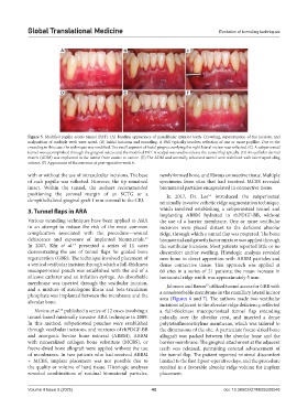
Figure 5. Modified papilla access tunnel (PAT). (A) Baseline appearance of mandibular anterior teeth. Crowding, supereruption of the incisors, and
malposition of multiple teeth were noted. (B) Initial incisions and tunneling. A PAT typically involves reflection of one or more papillae. Due to the
crowding in this case, the technique was modified. The small segment of facial gingiva overlying the right lateral incisor was reflected. (C) A subperiosteal
tunnel was accomplished through the gingival sulcus and the modified PAT. A scalpel was used to release the tunnel flap apically. (D) An acellular dermal
matrix (ADM) was implanted in the tunnel from canine to canine. (E) The ADM and coronally advanced tunnel were stabilized with interrupted sling
sutures. (F) Appearance of the outcome at post-operative week 6.
with or without the use of intrasulcular incisions. The base newly formed bone, and fibrous connective tissue. Multiple
of each papilla was reflected. However, the tip remained specimens from sites that had received MCBS revealed
intact. Within the tunnel, the authors recommended biomaterial particles encapsulated in connective tissue.
positioning the coronal margin of an SCTG or a In 2017, Dr. Lee introduced the subperiosteal
16
de-epithelialized gingival graft 1 mm coronal to the CEJ. minimally invasive esthetic ridge augmentation technique,
3. Tunnel flaps in ARA which involved establishing a subperiosteal tunnel and
implanting ABBM hydrated in rhPDGF-BB, without
Various tunneling techniques have been applied to ARA the use of a barrier membrane. One or more vestibular
in an attempt to reduce the risk of the most common incisions were placed distant to the deficient alveolar
complication associated with the procedure—wound ridge, through which a tunnel flap was prepared. The bone
33
dehiscence and exposure of implanted biomaterials. biomaterial and growth factor mixture was applied through
14
In 2007, Kfir et al. presented a series of 11 cases the vestibular incisions. Most patients reported little or no
demonstrating the use of tunnel flaps for guided bone discomfort and/or swelling. Histologic analysis revealed
regeneration (GBR). The technique involved placement of new bone in direct apposition with ABBM particles and
a vertical vestibular incision through which a full-thickness dense connective tissue. This approach was applied at
mucoperiosteal pouch was established with the aid of a 60 sites in a series of 21 patients; the mean increase in
silicone catheter and an inflation syringe. An absorbable horizontal ridge width was approximately 5 mm.
membrane was inserted through the vestibular incision, Johnson and Baron utilized tunnel access for GBR with
17
and a mixture of autologous fibrin and beta-tricalcium a nonabsorbable membrane in the maxillary lateral incisor
phosphate was implanted between the membrane and the area (Figures 6 and 7). The authors made two vestibular
alveolar bone.
incisions adjacent to the alveolar ridge deficiency, reflected
Nevins et al. published a series of 12 cases involving a a full-thickness mucoperiosteal tunnel flap extending
15
tunnel-based minimally invasive ARA technique in 2009. palatally over the alveolar crest, and inserted a dense
In this method, subperiosteal pouches were established polytetrafluoroethylene membrane, which was tailored to
through vestibular incisions, and mixtures of rhPDGF-BB the dimensions of the site. A particulate freeze-dried bone
and anorganic bovine bone mineral (ABBM), ABBM allograft was packed between the alveolar bone and the
with mineralized collagen bone substitute (MCBS), or barrier membrane. The gingival attachment at the adjacent
freeze-dried bone allograft were applied without the use teeth was released, permitting coronal advancement of
of membranes. In two patients who had received ABBM the tunnel flap. The patient reported minimal discomfort
+ MCBS, implant placement was not possible due to limited to the first 2 post-operative days, and the procedure
the quality or volume of hard tissue. Histologic analyses resulted in a favorable alveolar ridge volume for implant
revealed combinations of residual biomaterial particles, placement.
Volume 4 Issue 3 (2025) 42 doi: 10.36922/GTM025220048