
Page 53 - GTM-4-3
P. 53
Global Translational Medicine Evolution of tunneling techniques
A B C
D E F
G H I
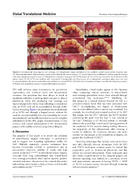
Figure 8. Circumferential tunneling for root coverage. (A) Subperiosteal tunnel established at the maxillary central incisor palatal recession sites.
(B) Tunneling instrument demonstrating a patent tunnel between the central incisors. (C) A facial tunnel was established to address gingival recession
at the right lateral and central incisors. (D) Subepithelial connective tissue graft (SCTG) harvested. (E) The SCTG was trimmed to the dimensions of the
palatal tunnel. (F) The SCTG was stabilized with interrupted 7-0 polyglycolic acid sling sutures. (G) A subpapillary continuous sling suture was used
to coronally advance the facial tunnel without the addition of a second SCTG. (H) Facial view of maxillary incisors 3 months following the procedure.
(I) Palatal view of maxillary incisors 3 months following the procedure.
IBD wall, enhance space maintenance for periodontal Nevertheless, mixed results appear in the literature
regeneration, and minimize facial and interproximal when comparing clinical outcomes of tunnel-based
recession. The procedure has been shown to result in root coverage procedures versus those attained through
significant reduction in probing depth and gain in clinical conventional flap techniques. 23,56-59 Stabilizing the
attachment, while also producing root coverage and flap margin in a coronal position beyond the CEJ is a
improved papilla fill. Rather than reflecting a conventional procedure-related factor that has been associated with
flap, an SCTG wall can be accomplished through multi- CRC. Accomplishing this degree of advancement
60
surface tunneling. Figure 9 demonstrates the establishment may be more difficult when a tunnel flap is utilized. For
of an SCTG wall through a lingual tunnel, without the example, the described post-surgical position of the PAT
12
need for conventional flap reflection. Extending the tunnel flap margin was the CEJ. Likewise, the MiTT involved
interproximally and facially permitted access for complete positioning the graft—not the flap—1 mm coronal to
13
debridement of the IBD, proper instrumentation of root the CEJ. Zabalegui et al. did not attempt to coronally
6
surfaces, and coronal advancement of the interproximal advance the tunnel flap, intentionally leaving portions of
gingiva, with stability achieved using a bone allograft. the implanted SCTG exposed during healing. However,
the magnitude of flap advancement after suturing is
6. Discussion known to influence the treatment outcome—the more
The purpose of this report is to review the evolution coronal the flap margin at the completion of surgery, the
of tunnel-based surgical techniques in periodontics greater the likelihood of CRC. 60
and to suggest new tunneling applications within the Tunneling techniques that include external incisions
field. Multiple minimally invasive techniques have may offer clinically relevant advantages. Both the PAT
become increasingly utilized in periodontics due to and VISTA techniques enhance access for tunnel flap
documented long-term stability of results and high preparation, graft or biomaterial placement, and flap
patient acceptance. 3,23,26,54,55 In periodontal plastic surgery, release for coronal advancement. These techniques may
clinicians have long acknowledged that tunnel-based also reduce surgical trauma to the marginal gingiva and
root coverage procedures often lead to superior patient- decrease operating time. Indeed, in a systematic review
oriented outcomes, characterized by reduced discomfort and meta-analysis, outcomes following VISTA + ADM
and swelling, faster return to daily activities, and favorable or SCTG surpassed results obtained through tunneling
early esthetic results. 10-13,28-32,54,55 without external incisions. 30
Volume 4 Issue 3 (2025) 45 doi: 10.36922/GTM025220048