
Page 51 - GTM-4-3
P. 51
Global Translational Medicine Evolution of tunneling techniques
A B C
D E F
G H I
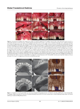
Figure 6. Tunnel access for guided bone regeneration (GBR). (A) Baseline appearance of maxillary anterior teeth. At the left central incisor position, a
substantial undercut in the alveolar bone could be palpated and appreciated visually. A cone-beam computed tomography scan confirmed inadequate
bone volume for implant placement. (B) Vertical vestibular incisions were placed in the midline frenum and between the left canine and lateral incisor. The
incisions extended from the depth of the vestibule 2–3 mm into the attached gingiva. A subperiosteal tunnel was established between the two vestibular
incisions. The full-thickness reflection extended palatally over the alveolar crest. A periosteal releasing incision was placed at the apical aspect of the tunnel.
(D) Intramarrow penetrations were established through the vertical incisions. (E) A dense polytetrafluoroethylene (PTFE) membrane was trimmed to
the dimensions of the site and inserted into the tunnel. A 4-0 chromic gut suture (arrow) needle was passed from the palatal aspect of the tunnel through
the midline vertical incision. The suture engaged the portion of the membrane designed to extend over the alveolar crest. (F) The needle was then passed
through the midline vertical incision, exiting the palatal mucosa adjacent to the original needle entry point (arrow). The membrane was then drawn into
place by exerting tension on both ends of the suture. (G) After implanting autogenous bone shavings and a freeze-dried bone allograft, the membrane was
stabilized with two fixation screws and an absorbable 5-0 polyglycolic acid mattress suture. (H) The vestibular incisions were closed with simple continuous
4-0 dense PTFE sutures. (I) Appearance of the site 6 months following GBR.
A B C
D E F
Figure 7. Comparison of baseline and follow-up cone-beam computed tomography volumes. (A) Baseline axial slice. (B) Baseline cross-sectional slice.
(C) Baseline volume rendering. (D) Axial slice 6 months following the procedure. (D) Cross-sectional slice 6 months following the procedure. (E) Volume
rendering 6 months following the procedure.
Volume 4 Issue 3 (2025) 43 doi: 10.36922/GTM025220048