
Page 48 - GTM-4-3
P. 48
Global Translational Medicine Evolution of tunneling techniques
A B C
D E F
G H I
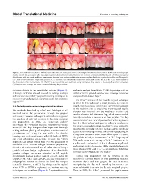
Figure 2. Coronally advanced tunnel with subepithelial connective tissue graft (SCTG). (A) Gingival recession defect <2 mm in depth at the maxillary right
central incisor. (B) Appearance after tunnel preparation between the two lateral incisors. (C) Coronal advancement of the tunnel. (D) After mechanical
debridement with ultrasonic and hand instruments, chemical root surface modification was accomplished with tetracycline hydrochloride (50 mg/mL).
(E) Close-up view of tunnel preparation prior to SCTG insertion. (F) Subepithelial connective tissue graft harvest site. (G) The SCTG was trimmed to
the dimensions of the recipient site. (H) The SCTG and tunnel flap were stabilized using interrupted sling sutures (4-0 dense polytetrafluoroethylene).
(I) Complete root coverage was noted 9 months following the procedure.
recession defects in the mandibular anterior (Figure 3). and meta-analysis found that a VISTA flap design with an
Although controlled clinical research is lacking, multiple ADM or SCTG yielded superior root coverage outcomes
authors have successfully adapted tunneling techniques to compared with tunnel flaps. 30
root coverage and gingival augmentation in this anatomic Dr. Chao introduced the pinhole surgical technique
11
region. 26,28,29 in 2012. In this technique, a small incision, 2–3 mm in
2.2. Techniques incorporating external incisions length, was placed near the depth of the vestibule adjacent
to the recipient site. A specialized transmucosal papilla
The methods described by Allen and Zabalegui et al. elevator was inserted into the vestibular incision and
6
5
involved tunnel flap preparation through the gingival used to elevate a full-thickness flap, which was extended
sulcus only. However, subsequent authors have suggested laterally to include a minimum of four papillae. The flap
the addition of external incisions to facilitate recipient was maintained in a coronal position by implanting two to
site preparation. In 2011, Dr. Homayoun Zadeh four 2 × 12-mm absorbable porcine collagen membranes.
10
introduced the vestibular incision subperiosteal tunnel No sutures, surgical dressings, or adhesives were applied to
access (VISTA) flap. This procedure began with thorough maintain the coronal position of the flap, and the vestibular
scaling and root planing, odontoplasty to reduce cervical access incisions were permitted to heal without suturing. In
prominences and bring the root within the alveolar a long-term case series with a mean follow-up of 14.5 years,
housing, and root conditioning with 24% buffered EDTA the pinhole technique demonstrated a CRC frequency of
gel. Four substantive distinctions between the VISTA 78% and a mean root coverage of 94%. Furthermore, in
31
approach and earlier tunnel flaps were the placement of a split-mouth randomized clinical trial comparing clinical
vestibular access incisions to begin the tunnel preparation,
elevation of a subperiosteal tunnel rather than utilizing a and patient-centered outcomes of the pinhole technique to
partial-thickness design, implantation of an absorbable those attained with CAF + SCTG, no significant difference
32
porcine collagen membrane saturated in 0.3 mg/mL between the two treatments was found.
recombinant human platelet-derived growth factor-BB Tunnel flap preparation can be complex in the
(rhPDGF-BB) rather than an SCTG, and use of bonded 6-0 mandibular anterior region at sites presenting minimal
polypropylene sutures to advance the facial flap margins recession depth and thin gingiva. In such situations,
coronally. However, a VISTA flap design can be applied manipulating the flap with tunneling instruments can
when ADM or SCTG is implanted, and various suturing cause substantial trauma to the delicate marginal gingiva.
techniques can be utilized (Figure 4). A systematic review Thus, in 2020, Dr. Allen presented the papilla access
12
Volume 4 Issue 3 (2025) 40 doi: 10.36922/GTM025220048