
Page 23 - IJB-1-1
P. 23
Manyi Wang, Jiankang He, Yaxiong Liu, et al.
a pulsed laser source, a reservoir of cells (donor
container), and a building substrate onto which the
cells are deposited to for construction as shown in
Figure 1 [9,17] . It is now feasible to write multiple cell
types synchronously with a very high printing resolu-
tion at the micron scale [18,19] . The systems that use UV
or other light sources are normally defined as 3D pro-
jection stereolithography, in which a Digital Micro-
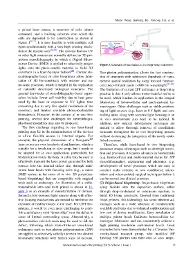
mirror Device (DMD) is applied to selectively project Figure 3. Schematic of laser-based in vivo bioprinting in dentistry.
lights onto the photo-curable material to build 3D
constructs in a layer-by-layer fashion [20] . Current ste- Two-photon polymerization allows for fast construc-
reolithography-based in vitro bioprinters allow fabri- tion of structures with submicron (hundreds of nano-
cation of 3D bio-constructs with micron- and na- meters) spatial resolution by using focused femtose-
no-scale precision, which is helpful in the replication cond near-infrared lasers (~800 nm wavelength) [21,22] .
of naturally developed biological structures. The The limitation of current 2PP technique in bioprinting
general drawbacks of stereolithography-based appro- practice is that it only allows mono-material resins to
aches include: lower cell viability due to heat gene- be used, which hinders its application in the integral
rated by the laser or exposure to UV lights; time fabrication of heterocellular and multi-material tis-
consuming due to very fine spatial resolutions of the sues/organs. Other challenges such as stable position-
construct; and limited available photocrosslinkable ing of light sources (e.g., laser or UV light) and con-
biomaterials. However, in the context of in vivo bio- trolling units, along with accurate light focusing in an
printing, several new challenges for stereolithogra- in vivo environment also need to be tackled. In
phy-based modalities may also be proposed. addition, new surgical debridement techniques are
The first challenge regarding in vivo laser-based needed to allow thorough removal of crosslinkers
printing may lie in the miniaturization of the devices remnants throughout the in vivo bioprinting process
to allow flexible access to internal organs. For without destroying the integration of the newly estab-
example, the physical dimensions of a Navigator TM lished construct.
laser source are over hundreds of millimeters, which is Therefore, while laser-based in vivo bioprinting
suitable for a bench-top in vitro setup, but it needs to possesses unique advantages such as ultrahigh resolu-
be adapted for in vivo application, particularly for tion and precision, it requires advances in biomaterials
biofabrication inside the body. A cable may be used to (e.g. heterocellular and multi-material resins for 2PP
effectively transmit the laser power generated by bulk stereolithography), engineering and photonics (e.g.
sources into the internal defect site, through mini- development of novel processes which are easy to
mized laser heads with focusing units (e.g., a micro conduct under extreme in vivo conditions), micro-
DMD system in the cases of in vivo 3D projection- robot- and robot-assisted surgical techniques before it
based bioprinting) that are compatible with surgical can be moved into clinical practices.
tools such as endoscopy. An illustration of a cable- (2) Inkjet-based bioprinting. Inkjet-based bioprinters
transmittable intra-oral teeth printer is shown in Fi- spray bioinks onto the deposition surface, either
gure 3 as an example of miniaturization of devices. through drop-on-demand or continuous ejection, to
Secondly, low-powered light sources along with effec- build 3D living constructs. Derived from traditional
tive focusing mechanisms are needed to minimize the inkjet printers, this technology has some inherent ad-
exposure of healthy tissues to the laser. For LIFT bio- vantages such as a wide selection of commercially
printers, it would be even more challenging to estab- available platforms due to technical sophistication and
lish a satisfactory mini “donor slide” over the defect in low cost of device modification. Easy installation of
cases of limited surrounding space. Alternatively, a multiple printer heads facilitates heterocellular tis-
photosensitive cell-rich resin may be used to fill the sue/organ fabrication and can concurrently achieve a
defect, following which state-of-the-art laser-based high printing resolution (sub-micron level). Good
techniques such as two-photon polymerization (2PP) examples have been demonstrated by a Clemson Uni-
are applied to selectively solidify the resin into desired versity-based research group, who modified HP
biomimetic structures with feature sizes of microns. Desktop 550 printers into their own in vitro inkjet-
International Journal of Bioprinting (2015)–Volume 1, Issue 1 19