
Page 171 - IJB-10-1
P. 171
International Journal of Bioprinting Bioprinted cell-laden hydrogel for tracheal application
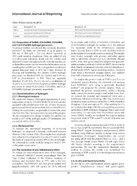
Table 1 Primers used in the qPCR
Gene Forward (5’–3’) Reverse (5’–3’)
TNF-α AGAACAGCAACTCCAGAACACCCT TGCCAGTTCCACATCTCGGATCAT
IL-6 AGACAGCCACTCACCTCTTCAG TTCTGCCAGTGCCTCTTTGCTG
GAPDH CGCTAACATCAAATGGGGTG TTGCTGACAATCTTGAGGGAG
2.2. Preparation of GelMA, ICA/GelMA, CS/GelMA, by co-culture with GelMA, ICA/GelMA, CS/GelMA, and
and ICA/CS/GelMA hydrogel precursors ICA/CS/GelMA hydrogels for another 24 h. We analyzed
To prepare GelMA, we followed the previously described the expression levels of the inflammatory cytokines
protocol. In brief, we dissolved 10 g of gelatin in tumor necrosis factor α (TNF-α) and interleukin 6 (IL-6)
18
500 mL of PBS (pH = 7.4) and stirred vigorously at in the samples by immunofluorescence staining. The samples
50°C until complete dissolution. Next, we added 10 mL were treated overnight with primary antibodies against
of methacrylate anhydride slowly into the solution and TNF-α (ab303458, Abcam) and IL-6 (ab183218, Abcam)
allowed it to react with gelatin for 2 h. After the reaction, we at 4°C. After rinsing, we treated the samples with goat anti-
collected the solution and removed insoluble substances by rabbit secondary antibody (1:1000, ab150077, Abcam) in the
centrifugation at 5000 rpm. The crude product was dialyzed dark. Finally, we mounted coverslips with 4′,6-diamidino-2-
against deionized water at 40°C for 3 days, followed by phenylindole (DAPI, Sigma) mounting solution, visualized
freezing and lyophilizing. For pristine GelMA hydrogel them using a fluorescent imaging system, and analyzed
precursors, we dissolved 10% w/v GelMA and 0.3% w/v them with a fluorescence microscope (Olympus).
LAP (a photoinitiator) in PBS. Then, we separately To analyze the protein levels of TNF-α and IL-6, we
dissolved 10 μM ICA, 2% w/v CS, and a combination of conducted western blotting. We extracted total proteins
10 μM ICA and 2% w/v CS in the pristine GelMA hydrogel from RAW264.7 cells using a previously described
precursors to obtain ICA/GelMA, CS/GelMA, and ICA/ method and prepared the protein samples. Then, we
30
CS/GelMA hydrogel precursors, respectively. measured the protein concentrations, added a loading
2.3. Characterizations of hydrogels buffer, mixed the protein samples, and boiled them. Next,
2.3.1. Rheological analyses we isolated the proteins and transferred them onto a
We conducted dynamic rheological experiments at room membrane, followed by blocking. After that, we immersed
temperature using the HAAKE MARS III photorheometer the protein strips in primary antibodies (1:1000, Beyotime,
equipped with parallel-plate (P20 TiL, 20 mm diameter) Shanghai, China) overnight at 4°C. After washing for 1 h,
geometry and OmniCure Series 2000 (365 nm, 20 mW/cm ). we incubated the strips with the corresponding secondary
2
During the experiments, we gradually increased the shear antibodies (1:2000; Beyotime, Shanghai, China) and rinsed
rate from 0 to 100 s to measure the viscosity of the samples. them thrice (15 min each) with Tris-buffered saline with
-1
We also determined the storage modulus (G’) and the loss tween (TBST). Finally, we developed the strips to analyze
modulus (G”) of the hydrogels under UV irradiation (365 the protein levels of TNF-α and IL-6, in which β-actin was
nm and 30 mW/cm ) for 400 s. used as control.
2
2.3.2. Mechanical test To analyze the gene expression levels of TNF-α and IL-6,
To evaluate the mechanical properties of cylindrical we carried out qPCR experiments. We extracted total RNA
hydrogels, we employed a dynamic mechanical analyzer using TRIzol™ reagent (Invitrogen) and performed cDNA
(Instron-5542; Canton, USA). The samples from all groups synthesis using Moloney murine leukemia virus reverse
were subjected to compression at a rate of 1 mm/min until transcriptase (Invitrogen). Quantification was carried
the depth of compression reached 30% of the initial height. out using qPCR with the primers listed in Table 1 and a
We computed the elastic modulus using the initial 0–20% Fast Synergy Brands Green Master Kit and Light Cycler
of the strain–stress curve. 480 System (Roche) according to the manufacturer’s
instructions. We used the comparative threshold cycle
2.4. The evaluation of anti-inflammatory effect method to analyze the results and normalized the
To evaluate the anti-inflammatory effect of the hydrogels, expression of TNF-α and IL-6 to the endogenous reference
we obtained RAW264.7 murine macrophages from the gene GAPDH.
Type Culture Collection of the Chinese Academy of
Sciences and incubated them at 37°C with a 5% CO 2.5. In vitro anti-bacterial activity
2
atmosphere. The RAW264.7 cells were pre-stimulated with To test the in vitro anti-bacterial activity against gram-
lipopolysaccharide (LPS, 100 ng/mL) for 24 h, followed positive (Staphylococcus aureus) and gram-negative
Volume 10 Issue 1 (2024) 163 https://doi.org/10.36922/ijb.0146