
Page 174 - IJB-10-1
P. 174
International Journal of Bioprinting Bioprinted cell-laden hydrogel for tracheal application
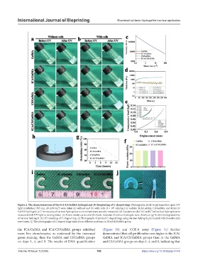
Figure 2. The characterizations of the ICA/CS/GelMA hydrogel and 3D bioprinting of C-shaped rings. Photographs of sol-to-gel transition upon UV
light irradiation (365 nm, 20 mW/cm ) were taken (a) without and (b) with cells (1 × 10 cells/mL) in GelMA, ICA/GelMA, CS/GelMA, and ICA/CS/
8
2
GelMA hydrogels. (c) The viscosity of various hydrogels at room temperature was also measured. (d) Dynamic moduli (G’ and G’’) of various hydrogels were
measured with UV light at varying times. (e) Stress–strain curves and (f) elastic modulus of various hydrogels were obtained. (g) In vitro biodegradability
of various hydrogels. (h) 3D modeling of C-shaped ring. (i) Photographs of printed C-shaped rings using various hydrogels pre-loaded with chondrocytes
were taken. (j) The photographs of C-shaped rings taken from different positions in ICA/CS/GelMA group.
the ICA/GelMA and ICA/CS/GelMA groups exhibited (Figure 5b) and CCK-8 assay (Figure 5c) further
more live chondrocytes, as evidenced by the increased demonstrated that cell proliferation was higher in the ICA/
green staining, than the GelMA and CS/GelMA groups GelMA and ICA/CS/GelMA groups than in the GelMA
on days 1, 4, and 9. The results of DNA quantification and CS/GelMA groups on days 1, 4, and 9, indicating that
Volume 10 Issue 1 (2024) 166 https://doi.org/10.36922/ijb.0146