
Page 177 - IJB-10-1
P. 177
International Journal of Bioprinting Bioprinted cell-laden hydrogel for tracheal application
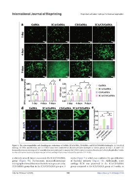
Figure 5. The cytocompatibility and chondrogenic evaluations of GelMA, ICA/GelMA, CS/GelMA, and ICA/CS/GelMA hydrogels. (a) Live/dead
staining, (b) DNA quantification, and (c) CCK-8 assay were conducted on chondrocyte-laden hydrogels in various groups on days 1, 4, and 9. (d)
Immunofluorescence staining and (e) quantification were performed to examine the COL II content in various chondrocyte-laden hydrogels after 3 weeks
in vitro cultivation. Statistical analysis was performed, and significance was indicated by asterisks (*P < 0.05).
a relatively smooth lumen was seen in the ICA/CS/GelMA weeks (Figure 7c), which was confirmed by quantification
group (Figure 7b). Furthermore, immunofluorescence of bacterial intensity (Figure 7d). Additionally, more
micrographs showed that more bacteria were present in the cartilage ECM was preserved in the ICA/CS/GelMA
ICA/GelMA group than in the ICA/CS/GelMA group at 3 group compared to the ICA/GelMA group at 3 weeks, as
Volume 10 Issue 1 (2024) 169 https://doi.org/10.36922/ijb.0146