
Page 411 - IJB-10-1
P. 411
International Journal of Bioprinting Droplets prepared by air-focused bioprinting
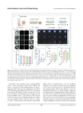
Figure 5. Encapsulation and controlled release of CAR-T cells in alginate hydrogel particles for immune therapy. (a) Schematics showing the encapsulation,
culture, release, and immune therapy of CAR-T cells in hydrogel particles. (b) Culture of CAR-T cells in hydrogel particles for 1, 3, 7, and 14 days. Cell
viability was characterized by live/dead staining kit. Live cells were stained in green by calcein AM, and dead cells were stained in red by PI. (c) Viability
of encapsulated cells cultured over time. Data are presented as mean ± SD (n = 8). (d) Bioluminescent images of Firefly luciferase-labeled AsPC-1 cells
after 2-day co-culture with (i) PBS, (ii) CAR-T cells encapsulated in hydrogel particles for 2 weeks and then released from hydrogel particles, and (iii)
unencapsulated CAR-T cells. (e) Relative luciferase intensity of firefly luciferase-labeled AsPC-1 cells after 2-day co-culture with released CAR-T cells and
free CAR-T cells. (f) IFN-γ and (g) IL-2 secreted by released CAR-T cells and unencapsulated CAR-T cells after 2-day co-culture. If not specified, data
were presented as mean ± SD (n = 6).
Pancreatic cancer cell line, firefly luciferase-labeled higher level of bioluminescence and thus luciferase
AsPC-1, which expressed a high level of MSLN, was level than those treated by released and unencapsulated
used as a model to test the immune therapy of MSLN- CAR-T cells, as shown in Figure 5e. In addition, released
targeting CAR-T cells, and the performances of CAR-T and unencapsulated CAR-T cells showed a good but
cells released from the hydrogel particles were investigated comparable performance in immune therapy, suggesting
by monitoring the cellular activity of cancer cells, e.g., the that encapsulation in hydrogel particles did not influence
level of luciferase. The bioluminescence images of firefly the CAR-T cell activity. The cell activity of released CAR-T
luciferase-labeled AsPC-1 cells treated by PBS released cells was further tested by measuring the levels of secreted
CAR-T cells after culture in hydrogel particles for 2 weeks, IFN-γ and IL-2, as shown in Figure 5f and g, respectively.
and unencapsulated CAR-T cells were shown in Figure Both IFN-γ and IL-2 secreted by released CAR-T cells after
5d. AsPC-1 cells treated by PBS showed a significantly culture in hydrogel particles for 2 weeks were comparable
Volume 10 Issue 1 (2024) 403 https://doi.org/10.36922/ijb.1102