
Page 458 - IJB-10-1
P. 458
International Journal of Bioprinting Efficacy of 3D-printed customized titanium implants
been registered on the website of Clinical Trials (www. surgical methods were subtalar arthrodesis, 3D-printed
clinicaltrials.gov; number: NCT03185286). Both patients customized titanium fusion cages, allograft cancellous
signed the informed consent form, agreeing to be enrolled implants, and internal fixation of screws (General Care,
in the study and disclose the case information. Both China). Postoperative procedures were the same as in
surgeries were performed by the same senior surgeon (X. Case 1.
Duan). Details of the surgical procedure and perioperative
complications were recorded, and regular follow-ups were 3. Results
conducted.
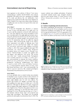
3.1. Surface morphology and microstructures
2.5.1. Case 1 The surface morphology, microstructure, and mechanical
A 57-year-old housewife was admitted to hospital properties between the 3D-printed plates and the screws
because of pain and deformity in the right ankle for prepared by traditional technology for TTC arthrodesis
15 years, and the symptoms worsened with claudication were compared and analyzed. From the appearance of the
for 1 year. Her diagnosis was severe osteoarthritis (OA) two implants (Figure 5), it could be seen that the screw had
and varus deformity of the right ankle and subtalar joints. been machined and its surface was smooth; the shape of
Using the aforementioned method, the 3D-printed ankle
joint models and implant samples were prepared based
on the CT examination data of the ankle for surgical
simulation. After confirming the details of the surgery,
three customized steel plates of different sizes were
prepared and then sterilized and sealed. The surgery
was successfully performed under epidural anesthesia
on May 11, 2016. The surgical methods were right TTC
arthrodesis and internal fixation using 3D-printed
customized plates and routine screws (General Care,
China). External fixation of plaster was applied after
the surgery, and antibiotics were given. The stitches
were taken out 2 weeks postoperatively. Radiographs
were routinely reviewed at 6 weeks, 12 weeks, and 1
year postoperatively to examine the fusion condition.
Blood tests were performed to check the blood routine
parameters, erythrocyte sedimentation rate (ESR),
C-reactive protein (CRP), liver and kidney functions,
etc. to comprehend the implant’s effects on body. After
the disease had been cured 1 year postoperatively, the Figure 5. Implants used in tibiotalocalcaneal (TTC) arthrodesis taken out
of the patients after fusion. (Left) 3D-printed customized plate for TTC
patient requested removal of the implant; pathological arthrodesis and (right) traditionally processed screws.
examination was performed in the local tissue.
2.5.2. Case 2
A 63-year-old lady, who is a retired worker, was admitted
to hospital because of pain associated with limitation of
motion of right hind foot for 2 years. Her diagnosis was
severe right subtalar joint OA with bone defects. The
patient had received left subtalar arthrodesis and iliac
bone grafting two years ago for similar pain in the left
hind foot. Although the left hind foot pain disappeared
after the surgery, the painful experience of taking the iliac
bone frightened her. Therefore, the patient decided to use
3D printing technology to treat the local bone defects.
Applying the similar 3D printing technique as in Case 1,
five titanium subtalar fusion cages were prepared and
sealed after sterilization. The surgery was successfully Figure 6. Surface morphology of customized plate for tibiotalocalcaneal
performed under epidural anesthesia on May 11, 2016. The (TTC) arthrodesis produced by electron beam 3D printing.
Volume 10 Issue 1 (2024) 450 https://doi.org/10.36922/ijb.0125