
Page 460 - IJB-10-1
P. 460
International Journal of Bioprinting Efficacy of 3D-printed customized titanium implants
Figure 8. Microstructure observation of customized plate by electron beam 3D printing (A, B) and screw by traditional methods (C, D). There were
obvious differences in the structures between the two implants. The inner part of the screw was small and even equiaxed structure; columnar crystals and
basket-weave microstructure were formed inside the customized plate. Different microstructures showed different mechanical properties. The hardness
results of the screw and the 3D-printed plate were 282.95 ± 2.22 HV and 327.50 ± 3.14 HV, respectively.
the patient had no recurrence of ankle deformity and the
follow-up examinations showed normal results (Figure 5).
The patient was satisfied with the treatment and willing to
recommend this new technology to her friends.
4. Discussion
4.1. Application of 3D printing in foot and ankle
surgery
The 3D printing is a type of rapid prototyping
technology, 22,25,26 which is based on digital model files and
uses powdery metal, plastics, or other adhesive materials
to construct objects with layer-by-layer printing. It is also
called additive manufacturing. In the past, it was often
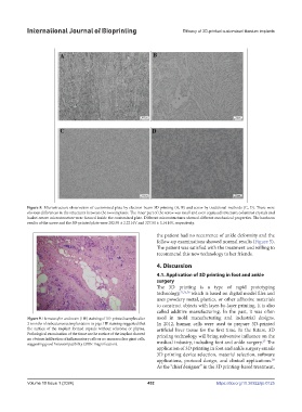
Figure 9. Hematoxylin and eosin (HE) staining of 3D-printed samples after used in mold manufacturing and industrial designs.
3 months of subcutaneous implantation in pigs. HE staining suggested that In 2012, human cells were used to prepare 3D-printed
the surface of the implant formed capsule without scleroma or phyma. artificial liver tissue for the first time. In the future, 3D
Pathological examination of the tissue on the surface of the implant showed printing technology will bring subversive influence on the
no obvious infiltration of inflammatory cells or no mononuclear giant cells, 27
suggesting good histocompatibility (200× magnification). medical industry, including foot and ankle surgery. The
application of 3D printing in foot and ankle surgery entails
3D printing device selection, material selection, software
applications, protocol design, and clinical applications.
28
As the “chief designer” in the 3D printing-based treatment,
Volume 10 Issue 1 (2024) 452 https://doi.org/10.36922/ijb.0125