
Page 457 - IJB-10-1
P. 457
International Journal of Bioprinting Efficacy of 3D-printed customized titanium implants
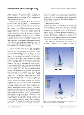
surgeons usually need to use 3D models and prefabricated When these conditions were determined, steel plate was
low-cost implants to perform surgical simulation for selected to solve the equivalent stress and equivalent elastic
preoperative planning, in order to take comprehensive strain (Figure 4). We demonstrated through finite element
surgical details into account. 5,9,20,21 simulation that the material had sufficient load-bearing
The procedure for preparing 3D-printed models and capacity during service.
implant samples were as follows. 22,23 Three-dimensional 2.5. Clinical applications
computed tomography (CT) scan (Siemens, Germany) of The inclusion criteria are as follows: (i) symptoms were
the ankle was routinely performed, and the CT scan slice obvious and did not relieve after more than 6 months of
thickness was 1 mm. The digital image correlation method conservative treatment; (ii) routine surgical treatment
(DICM) data were extracted and imported into the was flawed, and the patient refused to receive routine
MIMICS software to reconstruct the 3D data of the ankle surgery; and (iii) the patient had some knowledge of 3D
and the surrounding tissues. The MIMICS-reconstructed printing technology and wanted to receive precise surgical
data were then imported into the SIEMENS NX (Siemens, treatment. The exclusion criteria are as follows: (i) the
Germany) 3D design software. The engineer communicated patient could not be regularly followed; (ii) children and
with the surgeon to design the implant according to the pregnant women; and (iii) surgical contraindications.
surgical purpose. The designed model and the implant data
were converted into STL format and imported into a 3D This study was approved by the medical ethics
printer (model UP BOX, Tiertime, China). Polylactic acid committee (2016-J-001). The research protocol has
was used as raw materials for 3D printing.
In order to imitate the stress state after implantation,
finite element simulation was performed. The DICM data
of the 3D foot and ankle model were imported into the
Geomagic Studio Software (v2013, 3D system Inc., USA).
Grid command was used for smoothing and polishing the
surface. The 3D models of the screws and plates were also
imported into the Geomagic Studio Software. IGES format
of the 3D foot and ankle model was saved and imported
into the SIEMENS NX 12.0 3D design software. Stitch
command was used for integrating the model parts into
a coherent whole. XT format of the ensemble model was
saved and imported into the ANSYS Workbench software
(2021R1, ANSYS.USA), and static analysis module was
selected. Relevant parameters, including bone, cartilage,
steel plate, and screw material, were edited. Young’s
modulus parameters were set at 7300 MPA, 5 MPA,
120,000 MPA, 200,000 MPA, respectively, and Poisson’s
ratio parameters were set at 0.3, 0.46, 0.3, 0.3, respectively.
The mesh used in this analysis has a cell size of 1 mm. The
contacts between components (between steel plate and
screw, between steel plate and bone, and between screw
and bone) were all bound contacts, i.e., components were
inseparable in both normal and tangential directions to
simulate pressurized preloading between components.
Friction contact was set between bones and between bones
and cartilage with a friction coefficient of 0.7. According
to the literature, the vertical force of approximately five
times the body weight of the person was applied on each
foot during balanced standing. In order to simulate the
24
actual activity of the ankle joint in daily mode, we fixed
and constrained the lower end of the calcaneus and applied
a force of 285 N vertically downward to the upper surface Figure 4. Three-dimensional finite element models. (A) Equivalent stress
of the tibia according to the body weight of the patient. model and (B) equivalent elastic strain model.
Volume 10 Issue 1 (2024) 449 https://doi.org/10.36922/ijb.0125