
Page 456 - IJB-10-1
P. 456
International Journal of Bioprinting Efficacy of 3D-printed customized titanium implants
Table 2. Main technical parameters of electron beam melting
of Ti-6Al-4V alloy
Baseplate temperature 730°C
Preheat conditions Preheat electron beam = 30–38 mA
Preheat scan speed = (1–1.3) × 10 mm/s
4
Melting conditions Melting electron beam = 20 mA
Melting scan speed = 4500 mm/s
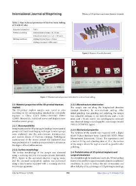
Figure 2. Diagram of model placement.
Figure 3. Titanium implants prepared with selective electron beam melting.
2.3. Material properties of the 3D-printed titanium 2.3.3. Microstructure observation
implant The sample was cut along the longitudinal direction
The 3D-printed implant samples were tested in vitro (vertical direction) by wire-electrode cutting. After
according to the corresponding standard for orthopedic initial grinding, fine grinding and polishing, the sample
implants in China (GB/T 16886.1-2011/ISO 10993- was etched by etchants (2 mL hydrofluoric acid + 4 mL
1:2009). Meanwhile, traditional screws and implants were nitric acid + 94 mL water). The metallographic structure
tested as control group. was observed using a metallographic microscope (model
PMG3, OLYMPUS, Japan).
2.3.1. Biocompatibility
Samples prepared by 3D printing technology (experimental 2.3.4. Mechanical properties
group) and traditional forging technique (control group) The hardness of the sample was measured with a digital
were implanted into the subcutaneous, intramuscular, micro Vickers hardness tester (model 401 MVD, Water
and osseous tissues of Guizhou minipigs. Pathological Measurement Instrument, China). The experiment used
examinations of the tissues around the implanted area a load of 0.3 kg and a loading time of 15 s. The surface
were performed at 3 months postoperatively to determine
the degree of local inflammation. of the sample should be kept as smooth as possible after
polishing.
2.3.2. Surface morphology
The surface morphology of the sample was observed 2.4. Prefabrication of 3D-printed implants and
using a scanning electron microscope (model JSM-6460, surgical simulation
JEOL, Japan) in the secondary electron imaging mode, As a breakthrough for traditional methods, 3D technology
and the chemical composition analysis was performed is now often used for surgical treatment of more complicated
using a spectrometer equipped with a scanning electron conditions. In order to reduce the risk of treatment and
microscope apparatus. allow full play of the advantages of 3D printing technology,
Volume 10 Issue 1 (2024) 448 https://doi.org/10.36922/ijb.0125