
Page 459 - IJB-10-1
P. 459
International Journal of Bioprinting Efficacy of 3D-printed customized titanium implants
the 3D-printed plate fabricated by electron beam could There was no fever or local immunological rejection
be customized according to the needs of the individual after operation, and the incision healed well 2 weeks
patient, but its surface was rough. Further analysis with postoperatively. Follow-up at 6 weeks showed normal
scanning electron microscopy showed that the plate by 3D results in ESR, CRP, blood routine parameters, and liver
printing method had more incompletely melted powder on and kidney functions. Radiographs showed satisfactory
the surface and obvious heaves due to fluid flow in the high- position of the TTC fusion as well as callus growth. The
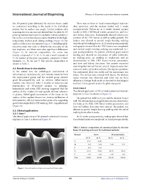
temperature molten pool during melting (Figure 6); the patient was allowed to partial weight-bearing walking
surface of the screw was smooth (Figure 7). Metallographic with the aid of walking boots. At 12 weeks postoperatively,
microstructure was used to observe the structure of the radiographs showed that the TTC fusion was completed,
two implants, and there were also significant differences and normal weight-bearing walking was conducted. At 1
(Figure 8). In material composition, the screw was year postoperatively, the patient exhibited good wound
mainly composed of Ti, Al, V, O, and a small amount of healing and showed no recurrence of ankle deformity,
P; the surface of the plate was mainly composed of three no walking pain, no swelling or tenderness, and no
elements, i.e., Ti, Al, and V. The specific composition is abnormalities in ESR, CRP, blood routine parameters,
shown in Table 3. and liver and kidney functions. The patient requested
removing the internal fixation, and all implants were then
3.2. Results from in vivo studies removed under epidural anesthesia. During the operation,
In the animal test, the pathological examination of the customized plate was stable and the appearance was
subcutaneous, intramuscular, and osseous tissues in both intact. The surface was covered with fascia. No obvious
the experimental group and the control group showed tissue reaction was observed, and there was no local
good biocompatibility, and no obvious inflammatory effusion or foreign body node as detected by histological
reaction was observed. After 3 months of subcutaneous method. Callus growth under the plate was good.
implantation of 3D-printed samples in minipigs,
hematoxylin and eosin (HE) staining suggested that the 3.3.2. Case 2
surface of the implant formed capsule without scleroma The clinical application of 3D-printed customized titanium
or phyma. Pathological examination of the tissue on the implants in Case 2 is shown in Figure 11.
surface of the implant showed no obvious infiltration of The patient had mild local pain and the incision healed
inflammatory cells nor mononuclear giant cells, suggesting well. No obvious immunological rejection was observed in
good histocompatibility (HE staining, 200× magnification) the body, as the ESR, CRP, blood routine parameters, and
(Figure 9). liver and kidney functions were normal. The patient was
3.3. Clinical applications allowed to partial weight-bearing walking with the aid of
3.3.1. Case 1 walking boots after 6 weeks.
The clinical application of 3D-printed customized titanium At 12 weeks postoperatively, radiographs showed that
implants in Case 1 is shown in Figure 10. the subtalar fusion was completed. At 1 year postoperatively,
Table 3. Surface composition of customized plate by electron
beam 3D printing and screw by traditional methods
Element Screw 3D-printed plate
wt, % at. % wt, % at. %
O 27.67 52.00
Al 5.22 5.81 5.69 9.71
P 0.53 0.52
Ti 63.17 39.66 88.72 85.24
V 3.41 2.01 5.59 5.05
Total 100.00 100.00 100.00 100.00
Figure 7. Smooth surface of the screw used with bone plate.
Volume 10 Issue 1 (2024) 451 https://doi.org/10.36922/ijb.0125