
Page 451 - IJB-10-1
P. 451
International Journal of Bioprinting Bioactive scaffold for necrosis bone repair
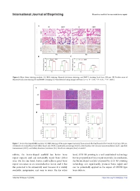
Figure 6. Bone tissue staining analysis. (A) H&E staining, Masson’s trichrome staining, and BMP-2 staining. Scale bars: 200 μm. (B) Positive areas of
Masson’s trichrome staining (B) and BMP-2 staining (C) were detected using ImageJ software (n = 6, *P < 0.05, **P < 0.01, ***P < 0.001).
Figure 7. In vivo biocompatibility analysis. (A) H&E staining of the major organs harvested from animals that had been fed for 8 weeks. Scale bars: 200 μm.
(B) Results of routine blood test (white blood cells [WBC], neutrophil percentage [NEU]), liver function test (alanine aminotransferase [ALT], aspartate
transaminase [AST]), and kidney function (creatinine [Cr], blood urea nitrogen [Bun]) at week 8.
rabbits, the biotin-doped scaffold has better bone hand, LTD 3D printing is a well-established technology
repair capacity and can sustainably repair bone defect for the preparation of bone repair materials. In conclusion,
area. On the one hand, biotin could achieve good bone the biotin-doped scaffold prepared by LTD 3D printing
repair outcomes as an osteoinductive factor, and it has technology can significantly promote bone repair and
the potential to be extensively used because it is widely can be potentially applied in the repair of ONFH-type
available, inexpensive, and easy to store. On the other bone defects.
Volume 10 Issue 1 (2024) 443 https://doi.org/10.36922/ijb.1152