
Page 446 - IJB-10-1
P. 446
International Journal of Bioprinting Bioactive scaffold for necrosis bone repair
of variance (ANOVA) and Tukey’s post hoc test. *P < 0.05, a microporous appearance. We used PLGA and β-TCP
**P < 0.01, and ***P < 0.001 were considered significant, as printing bioinks, so the elemental surface mapping of
and P > 0.05 was considered not statistically significant. the PT support (Figure S1 in Supplementary File) was
Statistical analyses were performed using SPSS 22.0 dominated by C, O, Ca, and P. However, biotin contains N
software (IBM, USA). and S elements, so we performed an elemental mapping of
the HBPT scaffold (Figure 1C) and found a comparatively
3. Results and discussion uniform distribution of N and S elements, indicating that
3.1. Preparation and characterization of scaffolds the scaffold was uniformly doped with biotin.
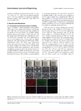
Figure 1A displays a camera photograph of the scaffold, FTIR was used to further validate the successful
which had a more regular and consistent morphology, doping of biotin in the scaffold. In FTIR (Figure 2A),
with visible ribs and macroscopically large openings for the stretching vibrations of O-H and N-H ranged from
-1
-1
the growth of new bone tissue. The scaffolds’ surfaces 3750 to 3000 cm . Thus, the peak at 3292 cm for the
appeared white because biotin is a white substance. The biotin powder is a stretching vibration of N-H and
low-magnification SEM image (60× magnification; Figure O-H, and the stretching vibration at 3009 cm for the
-1
1B) shows pore association with minor differences, as blank scaffold is O-H in PLGA. The remaining scaffold
well as curved prisms with a surface that was slightly contained biotin, and then N-H and O-H overlap at 3009
undulating. The high-magnification SEM image (2000× cm . Besides, biotin powder also showed characteristic
-1
magnification; Figure 1B) shows more pronounced peaks at 1277 cm , 1308 cm , 1640 cm , 1690 cm , and
-1
-1
-1
-1
microscopic pores on the surface of the PT scaffold, which 2923 cm , corresponding to C-O, C-N, N-H, C=O, and
-1
are special conduits that existed on the cryo-deposited C-H, respectively. The biotin-doped scaffolds suggested
3D-printed scaffold after lyophilization and promoted corresponding C-H, C-O, C-N/C-O, and C=O at 1092
cell adhesion and growth. Due to the presence of biotin in cm , 1191 cm , 1375 cm , and 1752 cm , respectively.
-1
-1
-1
-1
the LBPT, MBPT, and HBPT, the microscopic pores were In contrast, the blank scaffold only indicated C-O at
partially covered by biotin; however, as the biotin content 1375 cm . The above analysis and comparison revealed
-1
increased, the surface of the scaffold once again exhibited that the biotin powder was successfully doped in
Figure 1. Morphology of the material. (A) Top views of different scaffolds taken with a digital camera. (B) SEM images of the scaffolds at different
magnifications. Scale bars: 500 μm at 60× magnification, and 25 μm at 2000× magnification. (C) Elemental mapping analysis of the surface of the HBPT.
Scale bars: 25 μm.
Volume 10 Issue 1 (2024) 438 https://doi.org/10.36922/ijb.1152