
Page 450 - IJB-10-1
P. 450
International Journal of Bioprinting Bioactive scaffold for necrosis bone repair
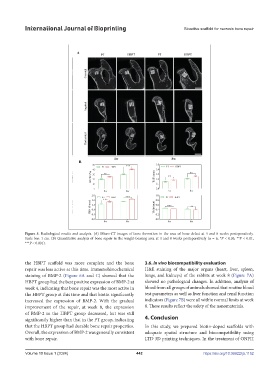
Figure 5. Radiological results and analysis. (A) Micro-CT images of bone formation in the area of bone defect at 4 and 8 weeks postoperatively.
Scale bar: 1 cm. (B) Quantitative analysis of bone repair in the weight-bearing area at 4 and 8 weeks postoperatively (n = 6, *P < 0.05, **P < 0.01,
***P < 0.001).
the HBPT scaffold was more complete and the bone 3.6. In vivo biocompatibility evaluation
repair was less active at this time. Immunohistochemical H&E staining of the major organs (heart, liver, spleen,
staining of BMP-2 (Figure 6A and C) showed that the lungs, and kidneys) of the rabbits at week 8 (Figure 7A)
HBPT group had the best positive expression of BMP-2 at showed no pathological changes. In addition, analysis of
week 4, indicating that bone repair was the most active in blood from all groups of animals showed that routine blood
the HBPT group at this time and that biotin significantly test parameters as well as liver function and renal function
increased the expression of BMP-2. With the gradual indicators (Figure 7B) were all within normal limits at week
improvement of the repair, at week 8, the expression 8. These results reflect the safety of the nanomaterials.
of BMP-2 in the HBPT group decreased, but was still
significantly higher than that in the PT group, indicating 4. Conclusion
that the HBPT group had durable bone repair properties. In this study, we prepared biotin-doped scaffolds with
Overall, the expression of BMP-2 was generally consistent adequate spatial structure and biocompatibility using
with bone repair. LTD 3D printing techniques. In the treatment of ONFH
Volume 10 Issue 1 (2024) 442 https://doi.org/10.36922/ijb.1152