
Page 447 - IJB-10-1
P. 447
International Journal of Bioprinting Bioactive scaffold for necrosis bone repair
scaffolds, and the characteristic functional groups of like structure and could maintain excellent compressive
biotin could be found in the FTIR spectra. Since the strength and drug release properties.
biotin was incorporated in trace amounts, there was
almost no difference between the FTIR spectra of these 3.2. In vitro evaluation of biocompatibility and
scaffolds. Subsequently, Figure 2B–D suggested that osteogenic properties of scaffold
the doping of biotin did not affect the porosity, pore Excellent biocompatibility of scaffold is a prerequisite
29,30
size, and filament diameter of the scaffold and that the for subsequent in vivo use of the scaffold. The results
of Live/Dead staining (Figure 3A) suggested that the
printed scaffold uniformly resembles the bionic structure rBMSCs proliferated well after 24 h and 72 h, and there
of cancellous bone, facilitating bone ingrowth. The were fewer dead cells (red) in all groups. CCK8 test (Figure
28
compressive test (Figure 2E) showed that all scaffolds 3B) yielded consistent results, indicating that biotin was
had cancellous bone-like compressive strength at the non-cytotoxic and did not affect cell proliferation. The
initial state, which decreased to varying degrees with adhesion of rBMSCs to the scaffold and the promotion of
increasing immersion time in simulated body fluid. The osteogenic differentiation of BMSCs by the scaffold were
HBPT scaffold maintained the best compressive strength subsequently observed in the co-culture of the scaffold
at week 8 due to the higher incorporation of biotin, and with rBMSCs. The ALP staining (Figure 3C) showed
all scaffolds showed mass loss with increasing immersion that all the biotin-doped scaffolds had better osteogenic
time (Figure 2F), but there was no significant difference effect than the normal scaffolds, with a larger area of
overall. Figure 2G shows that the release of biotin in the ALP-positive staining and a darker color. The HBPT
HBPT scaffold followed a rapid release in the first 3 weeks group achieved the best osteogenesis-related staining
(approximately 37.6% release), which may be due to the result because it contained more biotin. The results of the
fact that the surface area of the scaffold in contact with semi-quantitative analysis of ALP staining (Figure 3D)
body fluid is the greatest at the beginning. After 3 weeks, suggested that the HBPT group had the best osteogenic
the release of biotin plateaued, with approximately 62.4% effect on both day 6 and day 9 of osteogenic induction.
released at week 8. In summary, biotin-doped scaffolds can Cytoskeletal staining of rBMSCs on the scaffold (Figure
be successfully prepared by LTD 3D printing technology, 3E) suggested that the rBMSCs adhered well to the
and the structure of biotin could be preserved. The surface of the HBPT scaffold and that the cell pseudopods
prepared biotin-doped scaffolds held a cancellous bone- were spreading on the scaffold. Cytoskeleton staining at
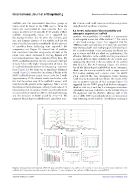
Figure 2. Characterization of materials. (A) FTIR spectrum of biotin powder and scaffolds. (B) Porosity, (C) pore size, and (D) filament diameter of the
scaffolds. (E) Compressive strength analysis of the scaffolds. (F) Mass variation of the scaffolds. (G) Biotin release curve in HBPT.
Volume 10 Issue 1 (2024) 439 https://doi.org/10.36922/ijb.1152