
Page 54 - IJB-2-1
P. 54
Patterning of tissue spheroids biofabricated from human fibroblasts on the surface of electrospun polyurethane matrix using…
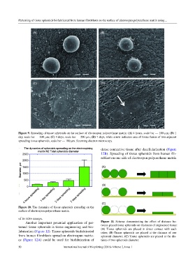
Figure 9. Spreading of tissue spheroids on the surface of electrospun polyurethane matrix: (A) 4 hours, scale bar — 100 µm; (B) 1
day, scale bar — 100 µm; (C) 4 days, scale bar — 200 µm; (D) 7 days, white arrow indicates area of tissue fusion of two adjacent
spreading tissue spheroids, scale bar — 100 µm. Scanning electron microscopy.
dense connective tissue after decellularization (Figure
12B). Spreading of tissue spheroids from human fib-
roblast on one side of electrospun polyurethane matrix
Figure 10. The dynamics of tissue spheroids spreading on the
surface of electrospun polyurethane matrix.
of in vitro assays.
Another important potential application of pat- Figure 11. Scheme demonstrating the effect of distance be-
terned tissue spheroids is tissue engineering and bio- tween placed tissue spheroids on thickness of engineered tissue:
(A) Tissue spheroids are placed in direct contact with each
fabrication (Figure 12). Tissue spheroids biofabricated other; (B) Tissue spheroids are placed at the distance of one
from human fibroblasts spread on electrospun matric- spheroid diameter; (C) Tissue spheroids are placed at the dis-
es (Figure 12A) could be used for biofabrication of tance of two spheroids diameter.
50 International Journal of Bioprinting (2016)–Volume 2, Issue 1