
Page 52 - IJB-2-1
P. 52
Patterning of tissue spheroids biofabricated from human fibroblasts on the surface of electrospun polyurethane matrix using…
Fusion of filaments with regular diameter leads to the
formation of larger diameter filaments. The electrospun
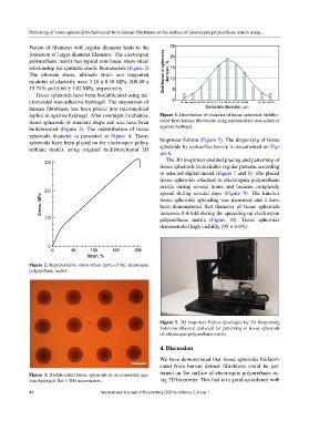
polyurethane matrix has typical non-linear stress-strain
relationship for synthetic elastic biomaterials (Figure 2).
The ultimate stress, ultimate strain and tangential
modulus of elasticity were 3.18 ± 0.48 MPa, 200.40 ±
15.74% and 6.66 ± 1.02 MPa, respectively.
Tissue spheroids have been biofabricated using mi-
cromolded non-adhesive hydrogel. The suspension of
human fibroblasts has been placed into micromolded
replica in agarose hydrogel. After overnight incubation, Figure 4. Distribution of diameter of tissue spheroids biofabri-
tissue spheroids of standard shape and size have been cated from human fibroblasts using micromolded non-adhesive
biofabricated (Figure 3). The redistribution of tissue agarose hydrogel.
spheroids diameter is presented at Figure 4. Tissue
spheroids have been placed on the electrospun polyu- bioprinter Fabion (Figure 5). The dispensing of tissue
rethane matrix using original multifunctional 3D spheroids by conus-like nozzle is documented on Fig-
ure 6.
The 3D bioprinter enabled placing and patterning of
tissue spheroids in desirable regular patterns according
to selected digital model (Figure 7 and 8). The placed
tissue spheroids attached to electrospun polyurethane
matrix during several hours and became completely
spread during several days (Figure 9). The kinetics
tissue spheroids spreading was measured and it have
been demonstrated that diameter of tissue spheroids
increases 8.4-fold during the spreading on electrospun
polyurethane matrix (Figure 10). Tissue spheroids
demonstrated high viability (95 ± 4.6%).
Figure 2. Representative stress-strain curve of the electrospun
polyurethane matrix.
Figure 5. 3D bioprinter Fabion developed by 3D Bioprinting
Solutions (Russia) and used for patterning of tissue spheroids
on electrospun polyurethane matrix.
4. Discussion
We have demonstrated that tissue spheroids biofabri-
cated from human dermal fibroblasts could be pat-
Figure 3. Biofabricated tissue spheroids in micromolded aga- terned on the surface of electrospun polyurethane us-
rose hydrogel. Bar = 200 micrometers. ing 3D bioprinter. This fact is in good accordance with
48 International Journal of Bioprinting (2016)–Volume 2, Issue 1