
Page 93 - IJB-2-1
P. 93
Wen Shing Leong, Shu Cheng Wu, Kee Woei Ng, et al.
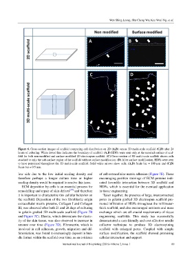
Figure 6. Cross-section images of scaffold comparing cell distribution on 2D (A,B) versus 3D multi-scale scaffold (C,D) after 24
hours of culturing. White dotted line indicates the boundary of scaffold. (A,B) HDFs were seen only at the external surface of scaf-
fold for both non-modified and surface modified 2D electrospun scaffold. (C) Cross-section of 3D multi-scale scaffold shows cells
attached to only the sub-surface region of the scaffold without surface modification. (D) After surface modification, HDFs were seen
to have penetrated throughout the 3D multi-scale scaffold. Solid white arrows show cells. (A,B) Scale bar = 100 µm and (C,D)
Scale bar = 0.5 mm.
low side due to the low initial seeding density and of cell-extracellular matrix adhesion (Figure 7E). These
therefore perhaps a longer culture time or higher encouraging positive stainings of ECM proteins indi-
seeding density would be required to resolve this issue. cated favorable interaction between 3D scaffold and
ECM deposition by cells is an essential process for HDFs, which is essential for the eventual application
remodelling and repair of skin defects [39] and therefore in tissue engineering.
it is important to characterize this cellular behavior on Taken together, the presence of large, interconnectted
the scaffold. Deposition of the two fibroblastic origin pores in gelatin grafted 3D electrospun scaffold pro-
extracellular matrix proteins, Collagen I and Collagen moted infiltration of HDFs throughout the millimeter-
III, was observed after both 21 and 28 days of culturing thick scaffold, and also encouraged nutrients and mass
in gelatin grafted 3D multi-scale scaffold (Figure 7B exchange which are all crucial requirements of tissue
and Figure 7C). Elastin, which determines the elastic- engineering scaffolds. This study has successfully
ity of the skin tissue, was also observed to increase in demonstrated a user-friendly and cost-effective needle
amount over time (Figure 7D). Fibronectin, which is collector technique to produce 3D electrospining
involved in cell adhesion, growth, migration and dif- scaffold with enlarged pores. Coupled with simple
ferentiation, was found to increasingly deposit in bun- surface modification, the scaffold showed promising
dle format within the scaffold over time, as an evidence cellular interaction and support.
International Journal of Bioprinting (2016)–Volume 2, Issue 1 89