
Page 92 - IJB-2-1
P. 92
Electrospun 3D multi-scale fibrous scaffold for enhanced human dermal fibroblasts infiltration
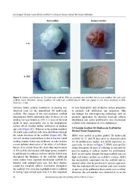
Figure 5. Surface modification on 3D multi-scale scaffold. PBS can penetrate into modified but not non modified 3D multi-scale
scaffold easily. Therefore, surface modified 3D multi-scale scaffold absorbs PBS and expand in size when immersed in PBS.
Scale bar = 2 mm.
collector, better cellular distribution at seeding was to have hydrophilic and conducive surface properties
observed even for the unmodified 3D multi-scale to promote cell infiltration and migration. This
scaffold. The images of the cryo-sectioned scaffold has bridged the electrospinning technology with its
demonstrated HDFs infiltration after 24 hours of cell potential application by showing thorough cellular
seeding but was limited to 21% (1.2 mm) of the total distribution and active proliferation into electrospun
depth (6 mm), presumably due to the hydrophobic scaffold with a thickness of a few millimeters.
surface which retarded further infiltration of medium
and cells (Figure 6C). Whereas in the gelatin modified 3.5 Gelatin Grafted 3D Multi-scale Scaffold for
3D multi-scale scaffold, cells were distributed through Dermal Tissue Engineering
the whole thickness of the scaffold (Figure 6D). The HDFs were seeded in gelatin grafted 3D multi-scale
low cell number observed here is due to the low initial scaffold for 21 and 28 days prior to characterization
seeding density which was deliberate in order to have for its proliferation marker and ECM deposition re-
a more definite observation of the effect of cell distri- spectively. As shown in Figure 7, HDFs were prolife-
bution. It is evident from this study that improvement rating throughout 28 days of culturing as indicated by
in 3D scaffold architecture with larger pores, wettabili- positive staining of cellular marker for proliferation,
ty and bioactivity enhance uniform cellular distribution Ki67, in cell nuclei. Despite the large scaffold size and
throughout the thickness of the scaffold. Although high cell density anchor on scaffold‘s surface, HDFs
some studies have reported electrospun scaffold fa- has successfully penetrated into the scaffold and re-
bricated with enlarged pore size, few have demon- mained viable with the proliferative protein expressed.
strated the complete cellular distribution throughout This has indicated efficient nutrient and mass transfer
the scaffold. Our study has highlighted that in addition in and out of the millimeter-thick multi-scale scaffold.
to having large enough pore size, it is also important However, the cell number was observed to be on the
88 International Journal of Bioprinting (2016)–Volume 2, Issue 1