
Page 94 - IJB-2-1
P. 94
Electrospun 3D multi-scale fibrous scaffold for enhanced human dermal fibroblasts infiltration
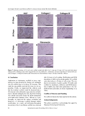
Figure 7. Histology staining of 3D multi-scale scaffold seeded with HDFs for 21 days and 28 days. Ki67 was positively stained
throughout the culturing period, which suggests that cells are proliferative. Furthermore, increased deposition of various ECM pro-
teins (Collagen I, Collagen III, Elastin, and Fibronectin) was observed from 21 days to 28 days. Scale bar = 200 µm.
4. Conclusion after 24 hours of cell seeding. Proliferation and ECM
deposition of HDFs in the gelatin grafted 3D scaffold
Application of electrospun scaffold in tissue engi- were observed up to 28 days of cell culture. The con-
neering has been hindered by limited cell infiltration ducive environment of gelatin grafted 3D multi-scale
into the scaffold due to its dense 2D structure. This scaffold for attachment, infiltration and ECM deposi-
study has successfully resolved this issue via two ap- tion of HDFs may find wide applications in the bio-
proaches. Firstly, we improvised the collector such medical field particularly in tissue engineering or as
that the resultant scaffold would be deposited into a fillers.
3D structure with sufficiently large pores for cell infil-
tration. Besides enlarged pore sizes, the fibers forming Conflict of Interest and Funding
3D scaffold mimicked closely the ECM architecture
with multi-scale diameters (from ~200 nm to 3 µm). No conflict of interest has been reported by the authors.
Secondly, we improved the surface wettability and Acknowledgements
bioactivity of electrospun scaffold through surface
modification. As a result, cell infiltration throughout The authors would like to acknowledge the support by
the entirety of the 6 mm fibrous scaffold was observed Nanyang Technological University.
90 International Journal of Bioprinting (2016)–Volume 2, Issue 1